. 2024 Aug 1:11:1443895.
doi: 10.3389/fnut.2024.1443895. eCollection 2024.

Omega-3 fatty acids abrogates oxido-inflammatory and mitochondrial dysfunction-associated apoptotic responses in testis of tamoxifen-treated rats

Affiliations

Omega-3 fatty acids abrogates oxido-inflammatory and mitochondrial dysfunction-associated apoptotic responses in testis of tamoxifen-treated rats

Adeyemi Fatai Odetayo et al. Front Nutr. .

Abstract

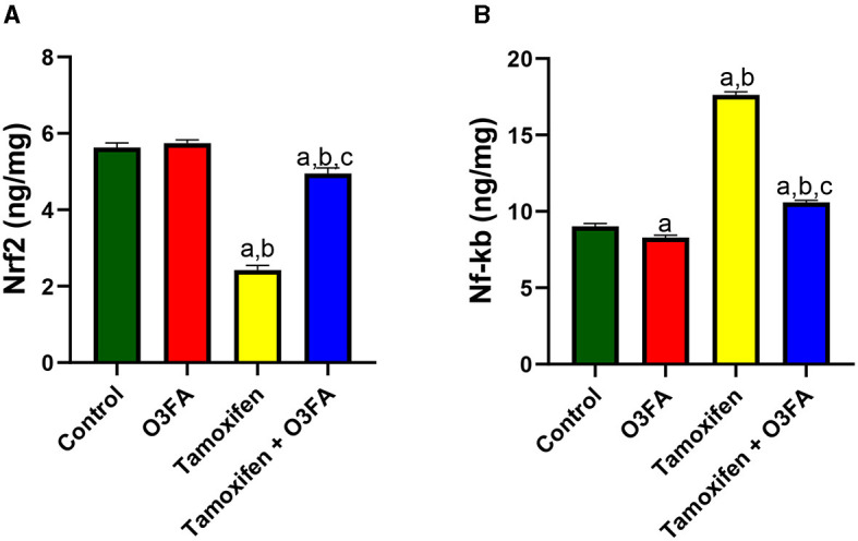
Background: Tamoxifen (TAM) is a widely used drug in patients with gynecomastia and breast cancer. TAM exerts its anticancer effects via its antiestrogenic activities. Unfortunately, TAM has been reported to exert gonadotoxic effects on male testes. Therefore, this study was designed to explore the possible associated mechanisms involved in TAM-induced testicular dysfunction and the possible ameliorative effects of omega-3 fatty acids (O3FA).

Methodology: Animals were randomly divided into control, O3FA, TAM, and TAM + O3FA. All treatment lasted for 28 days.

Results: TAM exposure impaired sperm qualities (count, motility, and normal morphology) and decreased testicular 3β-HSD and 17β-HSD. It was accompanied by a decline in serum testosterone and an increase in estradiol, luteinizing and follicle-stimulating hormones. These observed alterations were associated with an increase in testicular injury markers, oxido-inflammatory response, and mitochondria-mediated apoptosis. These observed alterations were ameliorated by O3FA treatments.

Conclusions: O3FA ameliorated TAM-induced testicular dysfunction in male Wistar rats by modulating XO/UA and Nrf2/NF-kb signaling and cytochrome c-mediated apoptosis in TAM-treated rats.

Keywords: anticancer drugs; cytochrome c; nuclear factor erythroid 2-related factor 2 (Nrf2) and nuclear factor-kappa B (NF-κB) signaling; selective estrogen receptor modulators; testicular function.


Conflict of interest statement

The authors declare that the research was conducted in the absence of any commercial or financial relationships that could be construed as a potential conflict of interest.

Figures

Figure 1
Figure 1
Effect of O3FA on sperm (A) count (B) motility (C) abnormal morphology in TAM exposed rats. aP < 0.05 vs. control, bP < 0.05 vs. O3FA; cP < 0.05 vs. TAM. Data were analyzed by one way ANOVA and Tukey's post-hoc test. O3FA, Omega-3 fatty acids; TAM, Tamoxifen.
Figure 2
Figure 2
Effect of O3FA on testicular (A) 3β-HSD (B) 17β-HSD in TAM exposed rats. aP < 0.05 vs. control, bP < 0.05 vs. O3FA; cP < 0.05 vs. TAM. Data were analyzed by one way ANOVA and Tukey's post-hoc test. O3FA, Omega-3 fatty acids; TAM Tamoxifen; 3β-HSD, 3 beta-hydroxysteroid dehydrogenase; 17β-HSD, 17 beta-hydroxysteroid dehydrogenase.
Figure 3
Figure 3
Effect of O3FA on serum (A) Luteinizing hormone (LH) (B) Follicle stimulating hormone (FSH) (C) testosterone (D) estradiol in TAM exposed rats. aP < 0.05 vs. control, bP < 0.05 vs. O3FA; cP < 0.05 vs. TAM. Data were analyzed by one way ANOVA and Tukey's post-hoc test. O3FA, Omega-3 fatty acids; TAM, Tamoxifen.
Figure 4
Figure 4
Testicular histology. Control, O3FA, and TAM + O3FA: section shows the testicular tissue composed of coils of seminiferous tubules (ST) with a defined lumen (L) containing sperm cells (SP), the seminiferous tubules contained germinal epithelium with germ cells at varying degree of maturation (line). The interstitium, contained blood vessel (arrow) which is free of collection and contained interstitial cells of Leydig (arrow head) appearing unremarkable. TAM, The blood vessels (black star) within the interstitium (plain star) appeared congested, and the interstitial cells of Leydig (arrow head) appears unremarkable.
Figure 5
Figure 5
Effect of O3FA on Johnsen Score in TAM exposed rats. aP < 0.05 vs. control, bP < 0.05 vs. O3FA; cP < 0.05 vs. TAM. Data were analyzed by one way ANOVA and Tukey's post-hoc test. O3FA, Omega 3 fatty acids; TAM, Tamoxifen.
Figure 6
Figure 6
Effect of O3FA on testicular (A) lactate (B) lactate dehydrogenase (LDH) (C) Gamma-glutamyl transferase (GGT) (D) sorbitol dehydrogenase (SDH) in TAM exposed rats. aP < 0.05 vs. control, bP < 0.05 vs. O3FA; cP < 0.05 vs. TAM. Data were analyzed by one way ANOVA and Tukey's post-hoc test. O3FA, Omega 3 fatty acids; TAM, Tamoxifen.
Figure 7
Figure 7
Effect of O3FA on testicular (A) malondialdehyde (MDA) (B) superoxide dismutase (SOD) (C) catalase (CAT) (D) glutathione (GSH) (E) glutathione peroxidase (GPx) (F) Glutathione-S-transferase (GST) in TAM exposed rats. aP < 0.05 vs. control, bP < 0.05 vs. O3FA; cP < 0.05 vs. TAM. Data were analyzed by one way ANOVA and Tukey's post-hoc test. O3FA, Omega 3 fatty acids; TAM, Tamoxifen.
Figure 8
Figure 8
Effect of O3FA on testicular (A) tumor necrotic factor-alpha (TNF-α) (B) interleukin 6 (IL-6) (C) myeloperoxidase (MPO) (D) nitric oxide (NO) in TAM exposed rats. aP < 0.05 vs. control, bP < 0.05 vs. O3FA; cP < 0.05 vs. TAM. Data were analyzed by one way ANOVA and Tukey's post-hoc test. O3FA, Omega 3 fatty acids; TAM, Tamoxifen.
Figure 9
Figure 9
Effect of O3FA on testicular (A) Xanthine oxidase (XO) (B) uric acid (UA) in TAM exposed rats. aP < 0.05 vs. control, bP < 0.05 vs. O3FA; cP < 0.05 vs. TAM. Data were analyzed by one way ANOVA and Tukey's post-hoc test. O3FA, Omega 3 fatty acids; TAM, Tamoxifen.
Figure 10
Figure 10
Effect of O3FA on testicular (A) Nrf2 (B) Nf-kb in TAM exposed rats. aP < 0.05 vs. control, bP < 0.05 vs. O3FA; cP < 0.05 vs. TAM. Data were analyzed by one way ANOVA and Tukey's post-hoc test. O3FA, Omega 3 fatty acids; TAM, Tamoxifen.
Figure 11
Figure 11
Effect of O3FA on testicular (A) cytochrome c (B) B-cell lymphoma 2 (BCl-2) (C) caspase 3 (D) DNA fragmentation index (DFI) in TAM exposed rats. aP < 0.05 vs. control, bP < 0.05 vs. O3FA; cP < 0.05 vs. TAM. Data were analyzed by one way ANOVA and Tukey's post-hoc test. O3FA, Omega 3 fatty acids; TAM, Tamoxifen.

Similar articles

Cited by

References

    1. Motrich RD, Ponce AA, Rivero VE. Effect of tamoxifen treatment on the semen quality and fertility of the male rat. Fertil Steril. (2007) 88:452–61. 10.1016/j.fertnstert.2006.11.196 - DOI - PubMed
    1. Kavishahi NN, Rezaee A, Jalalian S. The impact of miRNAs on the efficacy of tamoxifen in breast cancer treatment: a systematic review. Clin Breast Cancer. (2024) 24:341–50. 10.1016/j.clbc.2024.01.015 - DOI - PubMed
    1. De Oliveira Andrade F, Yu W, Zhang X, Carney E, Hu R, Clarke R, et al. . Effects of Jaeumkanghwa-tang on tamoxifen responsiveness in preclinical ER+ breast cancer model. Endocr Relat Cancer. (2019) 26:339–53. 10.1530/ERC-18-0393 - DOI - PMC - PubMed
    1. Senkoro E, Varadarajan M, Candela C, Gebreselassie A, Antoniadi C, Boffito M. Anastrozole as a therapeutic option for gynecomastia in a person receiving antiretroviral therapy: Case report. Br J Clin Pharmacol. (2024) 90:350–3. 10.1111/bcp.15951 - DOI - PubMed
    1. Sabanci E, Pehlivantürk-Kizilkan M, Akgül S, Derman O, Kanbur N. Tamoxifen treatment for pubertal gynecomastia: when to start and how long to continue. Breast Care. (2023) 18:249–55. 10.1159/000530408 - DOI - PMC - PubMed

LinkOut - more resources