. 2024 Jul 22:41:207-220.
doi: 10.1016/j.bioactmat.2024.06.030. eCollection 2024 Nov.

A functional analysis of a resorbable citrate-based composite tendon anchor

Affiliations

A functional analysis of a resorbable citrate-based composite tendon anchor

Arun Thirumaran et al. Bioact Mater. .

Abstract

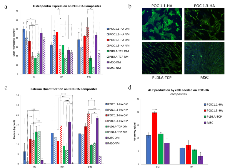
Rapid and efficient tendon fixation to a bone following trauma or in response to degenerative processes can be facilitated using a tendon anchoring device. Osteomimetic biomaterials, and in particular, bio-resorbable polymer composites designed to match the mineral phase content of native bone, have been shown to exhibit osteoinductive and osteoconductive properties in vivo and have been used in bone fixation for the past 2 decades. In this study, a resorbable, bioactive, and mechanically robust citrate-based composite formulated from poly(octamethylene citrate) (POC) and hydroxyapatite (HA) (POC-HA) was investigated as a potential tendon-fixation biomaterial. In vitro analysis with human Mesenchymal Stem Cells (hMSCs) indicated that POC-HA composite materials supported cell adhesion, growth, and proliferation and increased calcium deposition, alkaline phosphatase production, the expression of osteogenic specific genes, and activation of canonical pathways leading to osteoinduction and osteoconduction. Further, in vivo evaluation of a POC-HA tendon fixation device in a sheep metaphyseal model indicates the regenerative and remodeling potential of this citrate-based composite material. Together, this study presents a comprehensive in vitro and in vivo analysis of the functional response to a citrate-derived composite tendon anchor and indicates that citrate-based HA composites offer improved mechanical and osteogenic properties relative to commonly used resorbable tendon anchor devices formulated from poly(L-co-D, l-lactic acid) and tricalcium phosphate PLDLA-TCP.

Keywords: Citrate; Composite; Osteoinductive; Tendon anchor.


Conflict of interest statement

A single researcher who participated in this study was partly funded by Acuitive Technologies Ltd. The in vivo portion of this study was funded by a research grant from Acuitive Technologies Ltd.

Figures

Image 1
Graphical abstract
Fig. 1
Fig. 1
Uniaxial compression testing of POC-HA and PLDLA-TCP composites A) representative stress-strain curves, B) initial modulus, C) strain at stress, and D) peak stress. Data is represented as mean ± standard deviation. n = 4, * indicates p value of significance <0.01.
Fig. 2
Fig. 2
The effects of in vitro degradation on the surface roughness and mass loss of POC 1.1-HA, POC 1.3-HA and PLDLA-TCP substrates in vitro. Surface profilometry imaging (a) and subsequent analysis (b) of roughness (Sa) of PLDLA-TCP, POC 1.1-HA and POC 1.3-HA substrates maintained in PBS at for up to 14 days at 37 °C. Accelerated degradation analysis of mass loss of POC 1.1, POC 1.3 and PLDLA-TCP substrates immersed in PBS at 77 °C for up to 120 days (c).
Fig. 3
Fig. 3
DSC analysis of POC 1.1-HA, (a) POC 1.3-HA (b), and PLDLA-TCP (c), at 0, 7, and 14 days post-cell seeding. FTIR Spectra of POC 1.1-HA, POC 1.3-HA and PLDLA-TCP control substrates pre-cell seeding (day 0), and on day 7 and day 14 post-cell seeding (d).
Fig. 4
Fig. 4
Representative images of Calcein staining showing adhesion and growth of hMSCs on POC (a), POC 1.1-HA, (b), POC 1.3-HA (c) and PLDA-TCP (d). Scale bar 200 μm. Metabolic activity of hMSCs cultured on POC-HAs, PLDLA and tissue culture plastic for 7, 14 and 21 days normalized to cells in tissue culture plastic (B). hMSCs cultured on POC 1:1.3 composites demonstrated increased oxygen consumption rate relative to POC 1:1.1 and PLDLA-TCP control substrates (C). No significant differences in extracellular acidification rate were observed between any of the experimental or control groups (h). n = 3.
Fig. 5
Fig. 5
The in vitro osteospecific response of hMSCs cultured on POC-HA composites. Quantification of osteopontin expression (a) and immunostaining of osteopontin expression (b) in hMSC cultured on POC-HA and PLDLA-TCP composites in normal growth medium (NM) at day 21. Green osteocalcin, blue nucleus. (Magnification 20 ×, sale bar 100 μm. Calcium deposition (c) of hMSCs cultured on POC-HA and PLDLA-TCP composites in either osteogenic induction media (OM) or normal growth medium (NM) for 7, 14 and 21 days. Quantification of ALP activity measured on day 14 h in hMSCs cultured in OM (d).
Fig. 6
Fig. 6
Principle component analysis (PCA) plot for POC 1.1-HA, POC 1.3-HA, and PLDLA-TCP composites showing a clear separation between undifferentiated hMSCs grown in tissue culture plastic (MSC-NM), undifferentiated hMSCs grown on citrate-based composites and osteogenic differentiated hMSCs grown on tissue culture plastic (MSC-OM) (a). The number of differentially expressed genes for each group at each timepoint (b). Venn diagram showing unique and shared differentially expressed genes for all the composites at day 7 day 14 and day 21 (c). Ingenuity pathway analysis (IPA) was used to interpret differentially expressed genes biologically, generating a list of canonical pathways, diseases, functions, upstream regulators, and mechanistic networks. Pathways with a z score of more than 1.5 were considered significantly activated, while pathways with a z score of less than −1.5 were considered significantly inhibited. Detected enriched gene functions were plotted based on the significance of gene enrichment (-log P-value).
Fig. 7
Fig. 7
Analysis of the functional response of hMSCs to citrate composite materials. Heat map of osteo-responsive signaling pathways activated by POC 1.1-HA, POC 1.3-HA, and PLDLA-TCP (a) and heat map of enriched Bio-functions (b) leading to cytoskeletal rearrangement, cell migration, viability, blood vessel formation, extracellular matrix related functions, hMSC functions and lineage commitment and ossification related terms activated by citrate-based polymer composites (POC 1.1 and POC 1.3), and cells cultured on control PLDLA-TCP and tissue culture plastic substrates under osteogenic conditions. Plot based on activation score (Z value). A positive Z value represents activation of the biological function, while a negative Z value represents inhibition of the function. Analysis was performed relative to hMSCs cultured on tissue culture plastic under normal growth media conditions.
Fig. 8
Fig. 8
MicroCT and histological analysis of POC-HA and PLDLA-TCP bone anchor implants. MicroCT images depict the implant inside the tissue cavity, scale bar: 2 mm (a). Quantification of peri-implant tissue integration. significant reduction in peri-implant void was observed at 36 months post-implantation of citrate-HA tendon anchor devices (b), however, no significant differences were observed between groups at 6 months post-implantation (c). Histological analysis of the peri-implant region at 12 months post-implantation revealed that the presence of soft tissue was reduced and replaced by bone tissue (d). Ultimate load of Acuitive and Control treatments at the three time points. The “box” is bounded by the first and third quartiles; the “whiskers” represent the maximum/minimum values within the data set; the median data bar, mean data ‘x’, and outliers ‘·’ are highlighted. There were no significant differences between treatments (p = 0.910), timepoints (p = 0.100), or treatment-timepoint interactions (p = 0.710). Statistical significance of p < 0.05 is indicated *.

Similar articles

References

    1. Albrektsson T., Johansson C. Osteoinduction, osteoconduction and osseointegration. Eur. Spine J. 2001;10(Suppl 2):S96–S101. - PMC - PubMed
    1. Zhou Y., Hutmacher D.W., Varawan S.L., Lim T.M. In vitro bone engineering based on polycaprolactone and polycaprolactone–tricalcium phosphate composites. Polym. Int. 2007;56(3):333–342.
    1. Lohfeld S., Cahill S., Barron V., McHugh P., Dürselen L., Kreja L., Bausewein C., Ignatius A. Fabrication, mechanical and in vivo performance of polycaprolactone/tricalcium phosphate composite scaffolds. Acta Biomater. 2012;8(9):3446–3456. - PubMed
    1. Shao X., Goh J.C., Hutmacher D.W., Lee E.H., Zigang G.E. Repair of large articular osteochondral defects using hybrid scaffolds and bone marrow-derived mesenchymal stem cells in a rabbit model. Tissue Eng. 2006;12(6):1539–1551. - PubMed
    1. Fini M., Giavaresi G., Aldini N.N., Torricelli P., Botter R., Beruto D., Giardino R. A bone substitute composed of polymethylmethacrylate and α-tricalcium phosphate: results in terms of osteoblast function and bone tissue formation. Biomaterials. 2002;23(23):4523–4531. - PubMed

LinkOut - more resources