

Endotracheal tube cuff position in relationship to the walls of the trachea: A retrospective computed tomography-based analysis
- PMID: 39149725
- PMCID: PMC11323902
- DOI: 10.4103/sja.sja_36_24
Endotracheal tube cuff position in relationship to the walls of the trachea: A retrospective computed tomography-based analysis
Abstract
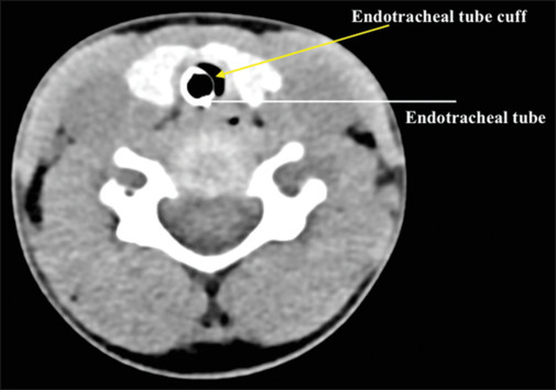
Background: The use of cuffed endotracheal tubes (ETTs) has become the standard of care in pediatric practice. The rationale for the use of a cuffed ETT is to minimize pressure around the cricoid while providing an effective airway seal. However, safe care requires that the cuff lie distal to the cricoid ring following endotracheal intubation. The current study demonstrates the capability of computed tomography (CT) imaging in identifying the position of the cuff of the ETT in intubated patients.
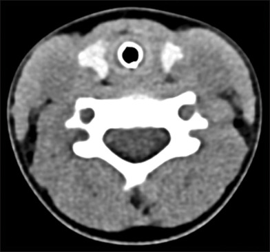
Methods: The study included patients ranging in age from 1 month to 10 years who underwent neck and chest CT imaging that required general anesthesia and endotracheal intubation. The location of the ETT and of the cuff within the airway was determined from axial CT images at three levels (proximal, middle, and distal). Anatomical orientations were tabulated, and percent chances of each orientation were determined for the ETT and the cuff.
Results: The study cohort included 42 patients ranging in age from 1 to 114 months. An ETT with a polyvinylchloride cuff was used in 24 patients, and an ETT with a polyurethane cuff was used in 18 patients. The ETT was located near the posterior wall of the trachea in approximately 24-38% of patients, being most likely to be centrally located at the proximal end and at its mid-portion. The middle part of the cuff was most likely to be positioned in the mid-portion of the trachea but tended to skew anteriorly at both the proximal and distal ends.
Conclusion: This is the first study using CT imaging to identify the uniformity of cuff inflation within the trachea in children. With commonly used cuffed ETTs, cuff inflation and the final position of ETT cuff within the tracheal lumen were not uniform. Future investigations are needed to determine the reasons for this asymmetry and its clinical implications.
Keywords: Computed tomography imaging; cricoid ring; endotracheal intubation; endotracheal tube cuff; pediatric airway; trachea.
Copyright: © 2024 Saudi Journal of Anesthesia.
Conflict of interest statement
There are no conflicts of interest.
Figures

References
-
- Khine HH, Corddry DH, Kettrick RG, Martin TM, McCloskey JJ, Rose JB, et al. Comparison of cuffed and uncuffed endotracheal tubes in young children during general anesthesia. Anesthesiology. 1997;86:627–31. - PubMed
-
- Motoyama EK. The shape of the pediatric larynx: Cylindrical or funnel shaped? Anesth Analg. 2009;108:1379–81. - PubMed
-
- Weiss M, Dullenkopf A, Fischer JE, Keller C, Gerber AC. Prospective randomized controlled multi-centre trial of cuffed or uncuffed endotracheal tubes in small children. Br J Anaesth. 2009;103:867–73. - PubMed
-
- de Wit M, Peelen LM, van Wolfswinkel L, de Graaff JC. The incidence of postoperative respiratory complications: A retrospective analysis of cuffed vs uncuffed tracheal tubes in children 0-7 years of age. Paediatr Anaesth. 2018;210:17–28. - PubMed
LinkOut - more resources
Full Text Sources