. 2024 Sep;33(9):e5144.
doi: 10.1002/pro.5144.

Structural basis for substrate flexibility of the O-methyltransferase MpaG' involved in mycophenolic acid biosynthesis

Affiliations

Structural basis for substrate flexibility of the O-methyltransferase MpaG' involved in mycophenolic acid biosynthesis

Cai You et al. Protein Sci. 2024 Sep.

Abstract

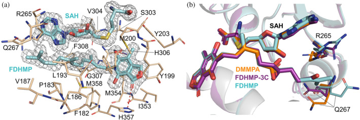
MpaG' is an S-adenosyl-L-methionine (SAM)-dependent methyltransferase involved in the compartmentalized biosynthesis of mycophenolic acid (MPA), a first-line immunosuppressive drug for organ transplantations and autoimmune diseases. MpaG' catalyzes the 5-O-methylation of three precursors in MPA biosynthesis including demethylmycophenolic acid (DMMPA), 4-farnesyl-3,5-dihydroxy-6-methylphthalide (FDHMP), and an intermediate containing three fewer carbon atoms compared to FDHMP (FDHMP-3C) with different catalytic efficiencies. Here, we report the crystal structures of S-adenosyl-L-homocysteine (SAH)/DMMPA-bound MpaG', SAH/FDHMP-3C-bound MpaG', and SAH/FDHMP-bound MpaG' to understand the catalytic mechanism of MpaG' and structural basis for its substrate flexibility. Structural and biochemical analyses reveal that MpaG' utilizes the catalytic dyad H306-E362 to deprotonate the C5 hydroxyl group of the substrates for the following methylation. The three substrates with differently modified farnesyl moieties are well accommodated in a large semi-open substrate binding pocket with the orientation of their phthalide moiety almost identical. Based on the structure-directed mutagenesis, a single mutant MpaG'Q267A is engineered with significantly improved catalytic efficiency for all three substrates. This study expands the mechanistic understanding and the pocket engineering strategy for O-methyltransferases involved in fungal natural product biosynthesis. Our research also highlights the potential of O-methyltransferases to modify diverse substrates by protein design and engineering.

Keywords: O‐methyltransferase; crystal structure; mycophenolic acid; protein engineering; substrate flexibility.


Conflict of interest statement

The authors declare no conflict of interest.

Figures

FIGURE 1
FIGURE 1
The biosynthetic steps mediated by the O‐methyltransferase MpaG'. The methyl groups installed by MpaG' are marked in red. The heads and tails of substrates are shaded in orange and blue, respectively.
FIGURE 2
FIGURE 2
Structural analysis of MpaG'/SAH/DMMPA complex. (a) Cartoon representation of the overall structure of MpaG'. The N‐terminal dimerization domain and C‐terminal catalytic domain are colored in yellow and pale green, respectively. The SAH and DMMPA molecules are both colored orange and shown as stick representations with a gray mesh illustrating the 2mF oDF c electron density map contours at the 1.0 σ level. (b) The dimeric state of MpaG'. One protomer is colored as presented in (a), and for the other protomer, the N‐terminal and C‐terminal domains are colored in blue and gray, respectively. (c) The enlarged view of the SAH binding pocket. The SAH and DMMPA molecules are presented as orange sticks. Hydrogen bonds and salt bridges are represented as red dashed lines. The distances between the sulfonium of SAH and the O1'/O2' of DMMPA are indicated by blue dashed lines. (d) The enlarged view of the DMMPA binding pocket, with hydrogen bond and salt bridge interactions indicated by red dashed lines.
FIGURE 3
FIGURE 3
HPLC analysis (254 nm) of the enzymatic reactions catalyzed by the wild type (WT) and mutant MpaG' enzymes using DMMPA (a), FDHMP‐3C (b), and FDHMP (c) as substrates. The analytical scale reactions containing 1 μM MpaG' (wild type or mutant), 5 mM SAM, and 0.5 mM substrate in 100 μl storage buffer were incubated at 40°C for 1 h.
FIGURE 4
FIGURE 4
Structural analysis of MpaG'/SAH/FDHMP‐3C complex. (a) The overall structure of the MpaG'/SAH/FDHMP‐3C complex. The N‐terminal and C‐terminal catalytic domains are colored pink and pale cyan, respectively. The enlarged view of SAH and FDHMP‐3C is shown as purple sticks with a gray mesh illustrating the 2mF oDF c electron density map contours at the 1.0 σ level. (b) Superimposition of the MpaG'/SAH/FDHMP‐3C complex and the MpaG'/SAH/DMMPA complex (gray). (c) Structural comparison of SAH binding pockets from MpaG'/SAH/FDHMP‐3C (SAH, purple; residues, pink) and MpaG'/SAH/DMMPA (SAH, orange; residues, gray). (d) The enlarged view of the FDHMP‐3C binding pocket, with hydrogen bond and salt bridge interactions indicated by red dashed lines. (e) Alternative conformations of the two residues R265 and Q267 in MpaG'/SAH/FDHMP‐3C (SAH, FDHMP‐3C, purple; residues, pale cyan) and MpaG'/SAH/DMMPA (SAH, DMMPA, orange; residues, gray).
FIGURE 5
FIGURE 5
Analysis of the MpaG'/SAH/FDHMP complex structure. (a) The binding pocket of FDHMP. The SAH and DMMPA molecules are shown in cyan sticks with a gray mesh illustrating the 2mF oDF c electron density map contours at the 1.0 σ level. Residues interacting with FDHMP are displayed as wheat sticks. Hydrogen bonds are represented as red dashed lines. (b) Structural alignment of three substrates DMMPA (orange), FDHMP‐3C (purple), and FDHMP (cyan) in the complex structures.
FIGURE 6
FIGURE 6
Structural comparison of MpaG' with other homolog proteins. (a) Structural alignment of MpaG' and the O‐methyltransferase‐like pericyclase LepI (PDB code: 6IX7). MpaG' is colored in yellow with helices α1 and α2 shown in orange and blue in the two monomers. LepI is shown in gray with helices α1 and α2 colored in cyan and light blue in the two monomers. (b) Structural alignment of MpaG' and classical OMT OxaC (PDB code: 5W7R). MpaG' is displayed in the yellow cartoon with helices α1 and α2 highlighted in orange. OxaC is displayed in cyan with helices α1 and α2 highlighted in marine. (c) Structural comparison of the substrate binding pockets from MpaG'/SAH/DMMPA (yellow) and LepI/SAH/ketone (gray). (d) Structural alignment of the substrate binding pockets from MpaG'/SAH/DMMPA (yellow) and OxaC/SAH/oxaline (pale cyan).
FIGURE 7
FIGURE 7
HPLC analysis (254 nm) of the enzymatic reactions catalyzed by the wild type (WT) and mutant MpaG' enzymes using DMMPA (a), FDHMP‐3C (b), and FDHMP (c) as substrates. The analytical scale reactions containing 1 μM MpaG' (wild type or mutant), 5 mM SAM, and 0.5 mM substrate in 100 μl storage buffer were incubated at 40°C for 1 h.

References

    1. Adams PD, Afonine PV, Bunkóczi G, Chen VB, Davis IW, Echols N, et al. PHENIX: a comprehensive python‐based system for macromolecular structure solution. Acta Crystallogr D Biol Crystallogr. 2010;66:213–221. - PMC - PubMed
    1. Bentley R. Mycophenolic acid: a one hundred year odyssey from antibiotic to immunosuppressant. Chem Rev. 2000;100:3801–3825. - PubMed
    1. Bernard SM, Akey DL, Tripathi A, Park SR, Konwerski JR, Anzai Y, et al. Structural basis of substrate specificity and regiochemistry in the MycF/TyIF family of sugar O‐methyltransferases. ACS Chem Biol. 2015;10:1340–1351. - PMC - PubMed
    1. Cai YJ, Hai Y, Ohashi M, Jamieson CS, Garcia‐Borras M, Houk KN, et al. Structural basis for stereoselective dehydration and hydrogen‐bonding catalysis by the SAM‐dependent pericyclase Lepl. Nat Chem. 2019;11:812–820. - PMC - PubMed
    1. Cullen D, Leong SA, Wilson LJ, Henner DJ. Transformation of Aspergillus nidulans with the hygromycin‐resistance gene, hph . Gene. 1987;57:21–26. - PubMed

MeSH terms

LinkOut - more resources