. 2024 Oct 15;92(10):e0030424.
doi: 10.1128/iai.00304-24. Epub 2024 Aug 16.

RSV enhances Staphylococcus aureus bacterial growth in the lung

Affiliations

RSV enhances Staphylococcus aureus bacterial growth in the lung

Helen E Rich et al. Infect Immun. .

Abstract

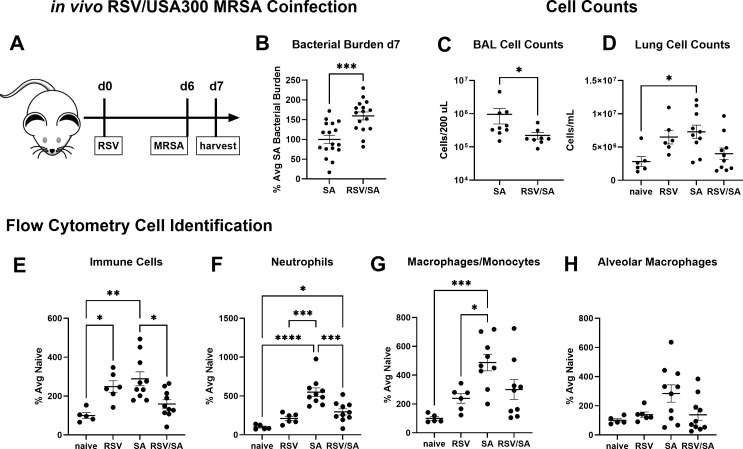
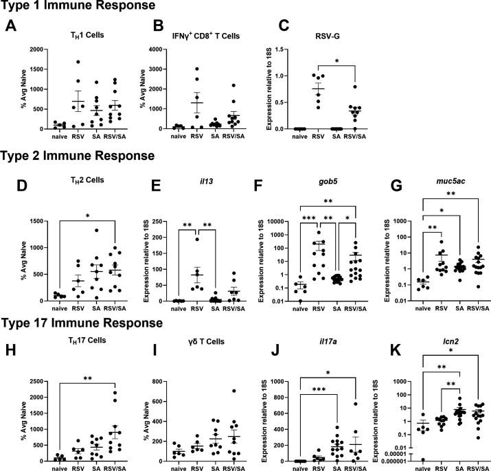
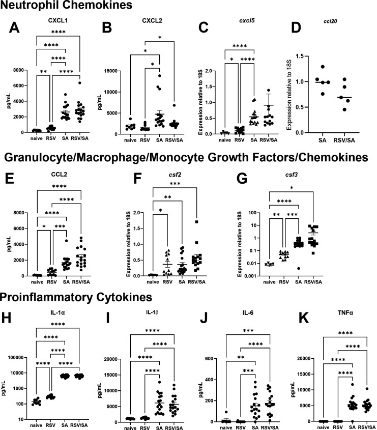
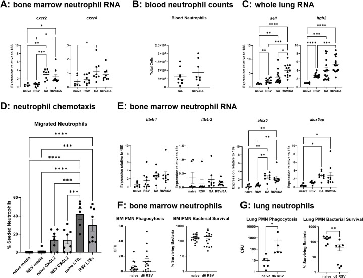
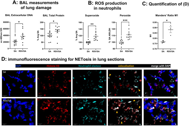
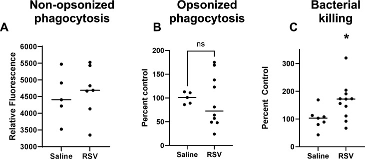
Patients coinfected with respiratory syncytial virus (RSV) and bacteria have longer hospital stays, higher risk of intensive care unit admission, and worse outcomes. We describe a model of RSV line 19F/methicillin-resistant Staphylococcus aureus (MRSA) USA300 coinfection that does not impair viral clearance, but prior RSV infection enhances USA300 MRSA bacterial growth in the lung. The increased bacterial burden post-RSV correlates with reduced accumulation of neutrophils and impaired bacterial killing by alveolar macrophages. Surprisingly, reduced neutrophil accumulation is likely not explained by reductions in phagocyte-recruiting chemokines or alterations in proinflammatory cytokine production compared with mice infected with S. aureus alone. Neutrophils from RSV-infected mice retain their ability to migrate toward chemokine signals, and neutrophils from the RSV-infected lung are better able to phagocytize and kill S. aureus ex vivo on a per cell basis. In contrast, while alveolar macrophages could ingest USA300 post-RSV, intracellular bacterial killing was impaired. The RSV/S. aureus coinfected lung promotes a state of overactivation in neutrophils, demonstrated by increased production of reactive oxygen species (ROS) that can drive formation of neutrophil extracellular traps (NETs), resulting in cell death. Mice with RSV/S. aureus coinfection had increased extracellular DNA and protein in bronchoalveolar lavage fluid and histological evidence confirmed NETosis in vivo. Taken together, these data highlight that prior RSV infection can prime the overactivation of neutrophils leading to cell death that impairs neutrophil accumulation in the lung. Additionally, alveolar macrophage killing of bacteria is impaired post-RSV. Together, these defects enhance USA300 MRSA bacterial growth in the lung post-RSV.

Keywords: MRSA; alveolar macrophage; lung; neutrophil; respiratory syncitial virus.


Conflict of interest statement

The authors declare no conflict of interest.

Figures

Fig 1
Fig 1
RSV rA2-line 19F inhibits USA300 MRSA clearance and limits neutrophil accumulation in the lung. (A) Schematic of mouse infections with harvest on day 7 post-RSV infection. (B) Mice were infected with RSV, challenged 6 days later with USA300 MRSA, and harvested 1 day after bacterial challenge. CFU from lung homogenate were plated on nutrient agar for enumeration. Data analyzed by Welch's t-test. (C) Cell counts from bronchoalveolar lavage or (D) collagenase digest of whole lung. Data analyzed by Mann-Whitney test (BAL Cell Counts) or by one-way Brown-Forsythe/Welch ANOVA (Lung Cell Counts). (E–H) Mice were infected, and lung cells were analyzed by flow cytometry as described in the methods. Data analyzed by one-way ANOVA (Immune Cells, all CD45+ cells, Neutrophils, Macrophages/Monocytes) or Brown-Forsythe/Welch ANOVA (Alveolar Macrophages). For ANOVAs, all comparisons were made. Asterisks are shown if differences are statistically significant (*P < 0.05, **P < 0.01, ***P < 0.005).
Fig 2
Fig 2
USA300 MRSA coinfection does not impair T cell responses to RSV. Mice were infected and lungs were processed via collagenase digest for flow cytometry, or all lung lobes were homogenized together and RNA was extracted from whole lung homogenate for real-time PCR as described in the methods. (A, B) TH1 and IFNγ-producing CD8 cell numbers were assessed by flow cytometry (compared by one-way ANOVA). (C) RSV glycoprotein expression was assessed by real-time PCR (analyzed by Welch's t-test as there was no expression detected in mice not infected with RSV). (D–G) TH2 cell numbers were assessed by flow cytometry, and il13, gob5, and muc5ac gene expressions were assessed by real-time PCR. Data were analyzed by one-way ANOVA (TH2 cells) or Kruskal-Wallis test (il13, gob5, muc5ac). (H–K) TH17 and γδ T cell numbers were assessed by flow cytometry, and il17a and lcn2 gene expressions were assessed by real-time PCR. Data were analyzed by one-way ANOVA (TH17 cells), Brown-Forsythe/Welch ANOVA (IFNγ+ CD8+ T cells), or Kruskal-Wallis test (il17a, lcn2). For ANOVAs and Kruskal-Wallis tests, all comparisons were made. Asterisks are shown if differences are statistically significant (*P < 0.05, **P < 0.01, ***P < 0.005).
Fig 3
Fig 3
Impaired bacterial clearance post-RSV infection is not explained by loss of cytokine or chemokine production. (A–D) Whole lung homogenate was assayed by ELISA for CXCL1 and CXCL2 protein, and cxcl5 or ccl20 gene expression relative to 18S was assayed in RNA extracted from whole lungs. Data for ccl20 are from a single experiment with n = 5. (E–G) Whole lung homogenate was assessed for CCL2 protein by ELISA, or RNA from whole lung was extracted and assayed by real-time PCR for expression of csf2 and csf3 relative to 18S. (H–K) Whole lung homogenate was assayed by ELISA for IL-1α, IL-1β, IL-6, and TNFα protein. All data were analyzed by one-way Brown-Forsythe/Welch ANOVA, with all comparisons made. Asterisks are shown if differences are statistically significant (*P < 0.05, **P < 0.01, ***P < 0.005).
Fig 4
Fig 4
RSV does not impair neutrophil antibacterial function or chemotaxis in vitro. (A) Primary bone marrow neutrophil RNA was extracted and assayed by real-time PCR for the expression of cxcr2 and cxcr4. All gene expression was analyzed relative to 18S and analyzed by one-way ANOVA. (B) Blood was harvested by cardiac puncture from mice infected with 2 × 10⁵ PFU RSV A2-line 19F or PBS control and challenged 6 days later with 5 × 10⁷ CFU of USA300 MRSA. The blood was assayed by flow cytometry for neutrophil numbers, analyzed by Welch's t-test. (C) RNA from whole lung was assayed for expression of sell and itgb2 and analyzed by Brown-Forsythe/Welch ANOVA. (D) Primary bone marrow neutrophils were assayed for chemotaxis toward LTB4, CXCL2, or media control and analyzed by one-way ANOVA. (E) Primary bone marrow neutrophil RNA was extracted and assayed by real-time PCR for the expressions of ltb4r1&2, alox5, and alox5ap. Data were analyzed by one-way ANOVA. (F) Neutrophils from bone marrow were assessed for bacterial uptake and killing analyzed by Mann-Whitney test. (G) Neutrophils from lungs were assessed for bacterial uptake and killing by bacterial plating and CFU determination analyzed by Mann-Whitney test. Asterisks are shown if differences are statistically significant (*P < 0.05, **P < 0.01, ***P < 0.005, ****P < 0.0005).
Fig 5
Fig 5
RSV infection preceding S. aureus challenge promotes lung damage and neutrophil hyperactivation. (A) BAL was obtained by lavage of the lung with 1 mL PBS from mice infected with 2 × 10⁵ PFU RSV A2-line 19F or PBS control and challenged 6 days later with 5 × 10⁷ CFU of USA300 MRSA. BAL supernatant collected on day 7 was assayed for extracellular DNA by sytox green staining and total protein by BCA assay. Data were analyzed by Welch's t-test. (B) Neutrophils from bone marrow of naïve or d6 RSV-infected mice were exposed to USA300 MRSA in vitro, then assayed for superoxide production by dihydroethidium oxidation assay or were assayed for peroxide production by Amplex Red assay. Data were analyzed by Welch's t-test. Asterisks are shown if differences are statistically significant (*P < 0.05, **P < 0.01, ***P < 0.005). (C) Mander's ratio quantification of colocalization of histone H-3 and neutrophil elastase staining in panel D. (D) Mice were infected with 2 × 10⁵ PFU RSV A2-line 19F or PBS control and challenged 6 days later with 5 × 10⁷ CFU of USA300 MRSA. Lung sections were stained with DAPI (blue) as well as antibodies to histone H3 (red) and neutrophil elastase (teal). Scale bar in lower left corner of each image corresponds to 10 µm. Orange arrowheads show areas of colocalization. Data in panel D are shown are from one mouse representative of three mice examined and quantified in panel C.
Fig 6
Fig 6
RSV infection impairs alveolar macrophage killing of S. aureus. Alveolar macrophages were harvested from saline or RSV-infected mice on day 6 by bronchoalveolar lavage. (A) Alveolar macrophages were plated with a 300:1 ratio of FITC-labeled USA300 MRSA and uptake was measured by intracellular fluorescence quenched extracellularly with trypan blue after 2 hours (data from one experiment and represents technical replicates of cells pooled from nine mice). (B) Alveolar macrophages were tested for phagocytosis of IgG-opsonized bacteria after 30 minutes. (C) Macrophages were allowed to kill bacteria ingested within 30 minutes over the next 90 minutes and were analyzed by CFU plating normalized to the mean of opsonized phagocytosis. Data in panels B and C are from two experiments combined and normalized to the saline control mean in each experiment as 100%. Data are technical replicates from n = 9 mice combined in each experiment and combined data were analyzed by student's t-test.

Similar articles

References

    1. Liu Y, Ling L, Wong SH, Wang MH, Fitzgerald JR, Zou X, Fang S, Liu X, Wang X, Hu W, Chan H, Wang Y, Huang D, Li Q, Wong WT, Choi G, Zou H, Hui DS, Yu J, Tse G, Gin T, Wu WK, Chan MT, Zhang L. 2021. Outcomes of respiratory viral-bacterial co-infection in adult hospitalized patients. EClinicalMedicine 37:100955. doi:10.1016/j.eclinm.2021.100955 - DOI - PMC - PubMed
    1. Falsey AR, Becker KL, Swinburne AJ, Nylen ES, Formica MA, Hennessey PA, Criddle MM, Peterson DR, Baran A, Walsh EE. 2013. Bacterial complications of respiratory tract viral illness: a comprehensive evaluation. J Infect Dis 208:432–441. doi:10.1093/infdis/jit190 - DOI - PMC - PubMed
    1. Crotty MP, Meyers S, Hampton N, Bledsoe S, Ritchie DJ, Buller RS, Storch GA, Micek ST, Kollef MH. 2015. Epidemiology, co-infections, and outcomes of viral pneumonia in adults: an observational cohort study. Medicine (Baltimore) 94:e2332. doi:10.1097/MD.0000000000002332 - DOI - PMC - PubMed
    1. Nguyen C, Kaku S, Tutera D, Kuschner WG, Barr J. 2016. Viral respiratory infections of adults in the intensive care unit. J Intensive Care Med 31:427–441. doi:10.1177/0885066615585944 - DOI - PubMed
    1. Glezen WP, Taber LH, Frank AL, Kasel JA. 1986. Risk of primary infection and reinfection with respiratory syncytial virus. Am J Dis Child 140:543–546. doi:10.1001/archpedi.1986.02140200053026 - DOI - PubMed

Publication types

MeSH terms

Substances

LinkOut - more resources