. 2024 Dec;16(4):976-989.
doi: 10.1007/s12539-024-00646-7. Epub 2024 Aug 16.

Luteinizing Hormone Receptor Mutation (LHRN316S) Causes Abnormal Follicular Development Revealed by Follicle Single-Cell Analysis and CRISPR/Cas9

Affiliations

Luteinizing Hormone Receptor Mutation (LHRN316S) Causes Abnormal Follicular Development Revealed by Follicle Single-Cell Analysis and CRISPR/Cas9

Chen Zhang et al. Interdiscip Sci. 2024 Dec.

Abstract

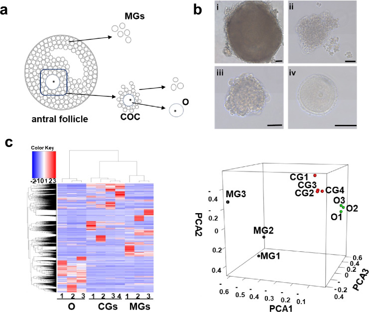
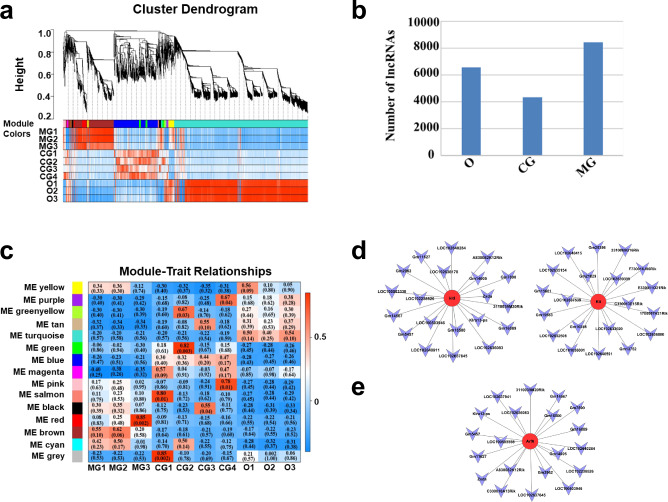
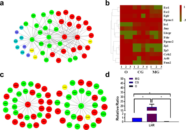
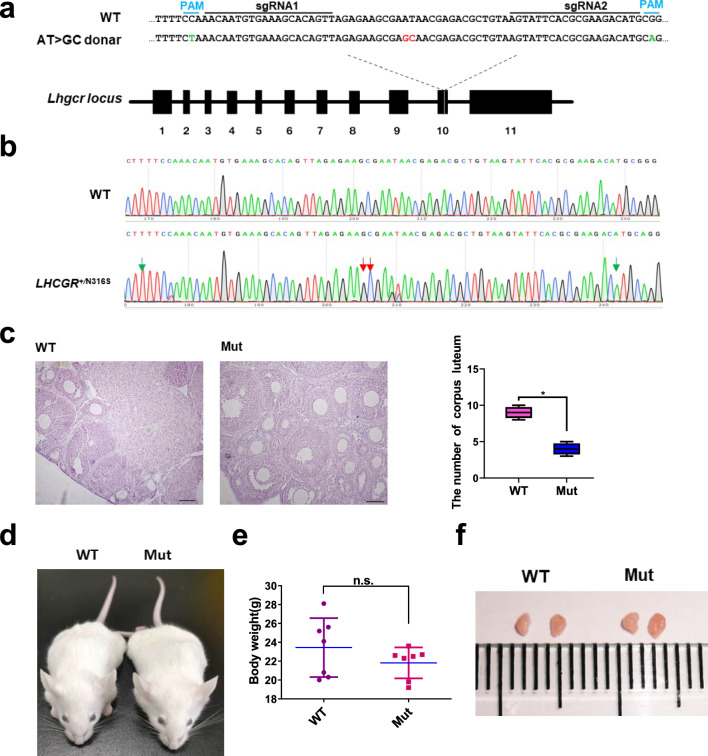
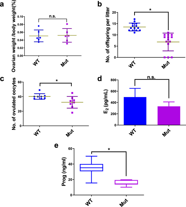
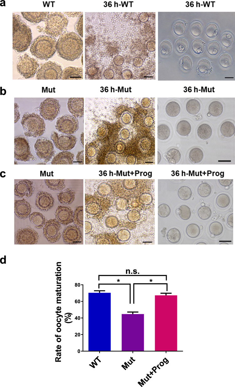
Abnormal interaction between granulosa cells and oocytes causes disordered development of ovarian follicles. However, the interactions between oocytes and cumulus granulosa cells (CGs), oocytes and mural granulosa cells (MGs), and CGs and MGs remain to be fully explored. Using single-cell RNA-sequencing (scRNA-seq), we determined the transcriptional profiles of oocytes, CGs and MGs in antral follicles. Analysis of scRNA-seq data revealed that CGs may regulate follicular development through the BMP15-KITL-KIT-PI3K-ARF6 pathway with elevated expression of luteinizing hormone receptor (LHR). Because internalization of the LHR is regulated by Arf6, we constructed LHRN316S mice by CRISPR/Cas9 to further explore mechanisms of follicular development and novel treatment strategies for female infertility. Ovaries of LHRN316S mice exhibited reduced numbers of corpora lutea and ovulation. The LHRN316S mice had a reduced rate of oocyte maturation in vitro and decreased serum progesterone levels. Mating LHRN316S female mice with ICR wild type male mice revealed that the infertility rate of LHRN316S mice was 21.4% (3/14). Litter sizes from LHRN316S mice were smaller than those from control wild type female mice. The oocytes from LHRN316S mice had an increased rate of maturation in vitro after progesterone administration in vitro. Furthermore, progesterone treated LHRN316S mice produced offspring numbers per litter equivalent to WT mice. These findings provide key insights into cellular interactions in ovarian follicles and provide important clues for infertility treatment.

Keywords: Follicle; Granulosa cells; LHRN316S; Oocytes; Progesterone; Single cell RNA-seq.


Conflict of interest statement

There is no disclosure of potential conflicts of interest.

Figures

Fig. 1
Fig. 1
Transcriptional profiles of single cells from the three types of cells examined in an antral follicle. a Schematic diagram of isolating individual cell types from a single antral follicle. b Morphology of a freshly isolated mouse antral follicle (i), mural granulosa cell (MG) (ii), cumulus-oocyte-complex (COC) (iii), and oocyte (O) (iv). c Unsupervised clustering of the transcriptome of all the samples. d Principal component analysis (PCA) of single cell expression patterns from the three type cells. Bar 50 μm
Fig. 2
Fig. 2
Gene-network modules established by weighted correlation network analysis (WGCNA), and oocyte polarity established gene-related long non-coding RNA (lncRNA). a WGCNA dendrogram indicating the expression of different gene modules in all 10 single-cell samples. b Module trait relationship followed by P values in parentheses between modules and different samples. c The number of lncRNAs detected. d and e LncRNAs correlated with genes Kitl, Kit and Arf6. Genes with different colored cycles mean different ranks in the network (ranks from higher to lower are red, green, yellow, dark blue and light blue). CG cumulus granulosa cells, O oocytes, MG mural granulosa cells
Fig. 3
Fig. 3
Gene-network and gene expression differences. a Genes co-expressed with Kit ligand (Kitl) and Kit in the network. b Genes co-expressed with Kitl in the network. Genes co-expressed with Kit in the network. c The gene expression of related genes in the three types of cells. d The validation of Lhr expression by qRT-PCR. O-oocytes, CG-cumulus granulosa cells, MG-mural granulosa cells. *P < 0.05, n.s. no significant difference
Fig. 4
Fig. 4
The generation of luteinizing hormone receptor (LHR)N316S mice. a Schematic view of the strategy used to generate the N316S point mutation (LHRN316S). Base pair substitutions for N316S are labeled in red. Silent mutations to prevent cleavage of the precisely mutated alleles are labeled in green. b Sanger sequencing chromatogram of genomic DNA from a wildtype (WT) mouse and the LHRN316S F0 founder. Red arrows indicate the base pair substitutions for the N316S point mutation. Green arrows indicate the silent mutations. c Morphological comparison of ovaries from LHRN316S and control female mice (left). The number of corpora lutea in different groups (right). Bar 100 μm. d The WT and LHRN316S C57BL/6 × ICR mice 8 weeks after birth. e The mean body weight of LHRN316S female mice was indistinguishable from that of controls (female littermates of LHRN316S female mice). f Images of whole ovaries from 8-week-old LHRN316S and control mice. *P < 0.05, n.s.-no significant difference
Fig. 5
Fig. 5
Low fertility of luteinizing hormone receptor (LHR)N316S adult female mice. a The relative ovarian weight of LHRN316S mice was indistinguishable from wild type (WT) mice. b Litter sizes from C57BL/6 × ICR (n = 14) LHRN316S female mice mated with ICR WT male mice were smaller than those from C57BL/6 × ICR (n = 14) WT female mice mated with ICR WT male mice. c The mean number of ovulated oocytes per female mouse after hormonal stimulation. d Estradiol (E2) levels in WT and LHRN316S mice. e Progesterone (Prog) homone levels in WT and LHRN316S mice. LHRN316S and WT adult female mice were used at the same ages. Data shown as mean ± SEM, n ≥ 6 mice per group. *P < 0.05, n.s.-no significant differences
Fig. 6
Fig. 6
Effect of progesterone on cumulus expansion and oocyte maturation. Cumulus oocyte complexes (COCs) were isolated from mice (48 h after pregnant mare serum gonadotropin stimulation) by puncturing large antral follicles and then cultured in IVM medium supplemented with or without 10 μmol/L progesterone for 36 h. a COCs from wild type (WT) mice were incubated in IVM medium. b COCs of luteinizing hormone receptor (LHR)N316S mice were incubated in IVM medium. c COCs from LHRN316S mice were incubated in IVM medium with 10 μmol/L progesterone. d The oocyte maturation rate of oocytes from LHRN316S mice incubated in IVM medium with or without progesterone. LHRN316S and WT female mice were used at the same ages. Bar 50 μm. *P < 0.05, n.s.-no significant differences

Similar articles

References

    1. Kidder GM, Vanderhyden BC (2010) Bidirectional communication between oocytes and follicle cells: ensuring oocyte developmental competence. Can J Physiol Pharmacol 88:399–413. 10.1139/y10-009 - PMC - PubMed
    1. Dupont J, Scaramuzzi RJ (2016) Insulin signalling and glucose transport in the ovary and ovarian function during the ovarian cycle. Biochem J 473:1483–1501. 10.1042/BCJ20160124 - PMC - PubMed
    1. Brunet S, Maro B (2007) Germinal vesicle position and meiotic maturation in mouse oocyte. Reproduction 133:1069–1072. 10.1530/REP-07-0036 - PubMed
    1. Barrett SL, Albertini DF (2010) Cumulus cell contact during oocyte maturation in mice regulates meiotic spindle positioning and enhances developmental competence. J Assist Reprod Genet 27:29–39. 10.1007/s10815-009-9376-9 - PMC - PubMed
    1. Gittens JE, Barr KJ, Vanderhyden BC et al (2005) Interplay between paracrine signaling and gap junctional communication in ovarian follicles. J Cell Sci 118:113–122. 10.1242/jcs.01587 - PubMed

LinkOut - more resources