. 2025 Feb;62(2):227-240.
doi: 10.1007/s00592-024-02349-3. Epub 2024 Aug 16.

Accumulation of branched-chain amino acids deteriorates the neuroinflammatory response of Müller cells in diabetic retinopathy via leucine/Sestrin2-mediated sensing of mTOR signaling

Affiliations

Accumulation of branched-chain amino acids deteriorates the neuroinflammatory response of Müller cells in diabetic retinopathy via leucine/Sestrin2-mediated sensing of mTOR signaling

Qiaoyun Gong et al. Acta Diabetol. 2025 Feb.

Abstract

Aims: This study aimed to investigate branched-chain amino acid (BCAA) catabolism in diabetic retinopathy (DR).

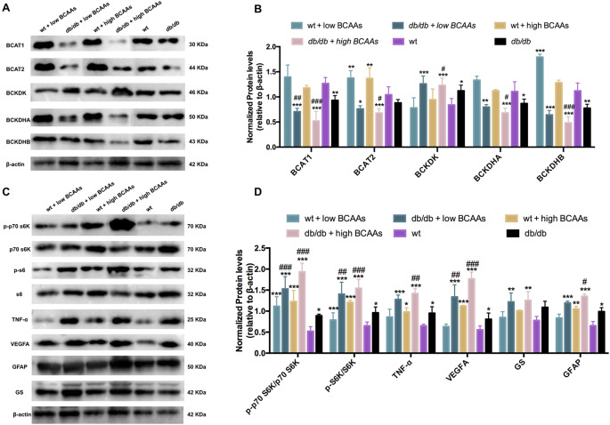
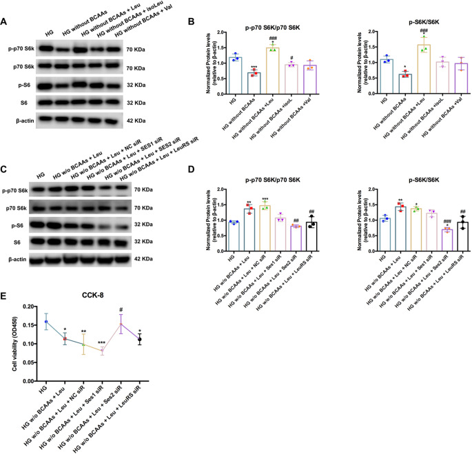
Methods: Wild-type and db/db mice were fed BCAAs (5 or 10 mg/kg/day) for 12 weeks, and hyperglycemia-exposed Müller cells were treated with BCAAs (2 or 5 mmol/L) for 24 and 48 h. BCAA levels were measured using MS/MS. Western blotting was performed to detect proteins. Flow cytometry, oxygen consumption rate, and Cell Counting Kit-8 assays were used to evaluate Müller cell viability. Each experiment was conducted at least thrice.

Results: BCAAs and branched-chain α-keto acids (BCKAs) were increased in the retina and systemic tissues of diabetic mice, and these changes were further enhanced to approximately 2-fold by extra BCAAs compared to wild-type group. In vitro, BCAAs and BCKAs were induced in hyperglycemic Müller cells, and augmented by BCAA supplementation. The aberrant BCAA catabolism was accompanied by mTORC1 activation and subsequently induced TNF-ɑ, VEGFA, GS, and GFAP in retinas and Müller cells under diabetic conditions. The cell apoptosis rate increased by approximately 50%, and mitochondrial respiration was inhibited by hyperglycemia and BCAA in Müller cells. Additionally, mTORC1 signaling was activated by leucine in Müller cells. Knockdown of Sestrin2 or LeuRS significantly abolished the leucine-induced mTORC1 phosphorylation and protected Müller cell viability under diabetic conditions.

Conclusions: We found that BCAA catabolism is hindered in DR through mTORC1 activation. Leucine plays a key role in inducing mTORC1 by sensing Sestrin2 in Müller cells. Targeting Sestrin2 may ameliorate the toxic effects of BCAA accumulation on Müller cells in DR.

Keywords: Branched-chain amino acids; Diabetic retinopathy; Glial; Inflammation; mTORC1.


Conflict of interest statement

Declarations. Ethics approval and consent to participate: All animal experiments were approved by the Animal Care and Use Committee of Shanghai First People’s Hospital of Shanghai Jiaotong University. Consent for publication: Not applicable. Competing interests: The authors declare that they have no competing interests.

Figures

Fig. 1
Fig. 1
Accumulated BCAAs and BCKAs in db/db mice fed with BCAAs and Müller cells. (A) The levels of BCAAs (leucine, isoleucine and valine) and BCKAs (KIC, KMV and KIV) were greater in the retinas of db/db mice than in those of wt mice. Food feeding with extra BCAAs further aggravated the high levels of BCAAs and BCKAs. The levels of BCAAs and BCKAs detected systemically in the plasma (B), kidney (C), liver (D) and heart (E). (F) In vitro, BCAAs and BCKAs were more abundant in high glucose-cultured Müller cells than in normal glucose-cultured Müller cells. The deprivation of BCAAs in high-glucose medium significantly suppressed the levels of BCAAs and BCKAs. The addition of a low dose or high dose of BCAAs for 24–48 h distinctly induced the accumulation of BCAAs and BCKAs. wt, wild type; db/db, diabetic mice; *p < 0.05, **p < 0.01, ***p < 0.001; n = 3
Fig. 2
Fig. 2
BCAA catabolism affected inflammation, neovascularization and glial activation in Müller cells via mTORC1 signaling in vivo. (A) The grey blots of enzymes involved in BCAA catabolism in the retinas of db/db mice fed with different doses of BCAAs. (B) Quantification of blots by ImageJ showed that BCAT1, BCAT2, BCKDHA and BCKDHB were inhibited in diabetic retinas and deteriorated by BCAAs supplementation, while BCKDK demonstrated the opposite changes in expression. (C) Western blots showing the phosphorylation of p70 S6K and S6K and TNF-α, VEGFA, GFAP and GS in the retinas. (D) Phosphorylation of p70 S6K and S6K was enhanced in the retinas of db/db mice and further increased by BCAAs feeding. The levels of TNF-α, VEGFA, GFAP and GS increased with BCAAs accumulation in diabetic retinas. wt, wild type; db/db, diabetic mice; *p < 0.05, **p < 0.01, ***p < 0.001, compared to the wt group; #p < 0.0, ##p < 0.01, ###p < 0.00, compared to the db/db group; n = 3. β-actin was used as the reference gene
Fig. 3
Fig. 3
Impaired BCAA catabolism contributed to cellular dysfunction in Müller cells under hyperglycemic conditions in vitro. (A) The grey blots of the BCAA catabolic enzymes in Müller cells treated with different doses of BCAAs under high glucose conditions for 24 h and 48 h. (B) Quantification of blots revealed that the protein levels of BCAT1, BCAT2, BCKDHA and BCKDHB were decreased in hyperglycemic cells and further inhibited by BCAAs addition, while BCKDK was enhanced significantly. (C, D) The expression levels of p-p70 S6K/p70 S6K, p-S6K/S6K, TNF-α, VEGFA, GFAP and GS were elevated by high glucose and BCAAs supplementation in Müller cells cultured for 24–48 h. (E-H) Müller cells were collected. The apoptotic rate and statistical analysis of Müller cells treated with different doses of BCAAs under HG conditions. (I, J) Mitochondrial dysfunction caused by high glucose and BCAAs accumulation in Müller cells was revealed by OCR and ATP production analysis (n = 4). *p < 0.05, **p < 0.01, ***p < 0.00, compared to the NG group; #p < 0.0, ##p < 0.01, ###p < 0.001, compared to the HG group; n = 3. GAPDH was used as the reference gene
Fig. 4
Fig. 4
Leucine activated mTORC1 signaling via Sestrin2/LeuRS in Müller cells under hyperglycemia. (A, B) Western blot experiments demonstrated that the phosphorylation of mTORC1 was significantly increased by leucine in Müller cells under hyperglycemia without complicated BCAAs. (C, D) Knockdown of Sestrin2 or LeuRS suppressed the stimulation of the mTORC1 complex, including p70 S6K and S6K, with leucine overexpression in Müller cells under high glucose conditions. (E) Sestrin2 interference protected cell viability from leucine-induced damage to Müller cells under HG conditions. *p < 0.05, **p < 0.01, ***p < 0.001, compared to the HG group; #p < 0.05, ##p < 0.0, ###p < 0.00, compared to the HG without BCAAs group for A and B, compared to the HG without BCAAs added leucine group for C and D; n = 3. β-actin was set as the reference gene

Similar articles

Cited by

References

    1. Hammes HP (2018) Diabetic retinopathy: hyperglycaemia, oxidative stress and beyond. Diabetologia 61(1):29–38 - PubMed
    1. Antonetti DA, Silva PS, Stitt AW (2021) Current understanding of the molecular and cellular pathology of diabetic retinopathy. Nat Reviews Endocrinol 17(4):195–206 - PMC - PubMed
    1. Bianco L, Arrigo A, Aragona E, Antropoli A, Berni A, Saladino A et al (2022) Neuroinflammation and neurodegeneration in diabetic retinopathy. Front Aging Neurosci 14:937999 - PMC - PubMed
    1. Rolev KD, Shu XS, Ying Y (2021) Targeted pharmacotherapy against neurodegeneration and neuroinflammation in early diabetic retinopathy. Neuropharmacology 187:108498 - PubMed
    1. Yang S, Qi S, Wang C (2022) The role of retinal Müller cells in diabetic retinopathy and related therapeutic advances. Front cell Dev Biology 10:1047487 - PMC - PubMed

MeSH terms

LinkOut - more resources