. 2024 Oct 15;30(20):4743-4754.
doi: 10.1158/1078-0432.CCR-24-0361.

YAP1 Status Defines Two Intrinsic Subtypes of LCNEC with Distinct Molecular Features and Therapeutic Vulnerabilities

Affiliations

YAP1 Status Defines Two Intrinsic Subtypes of LCNEC with Distinct Molecular Features and Therapeutic Vulnerabilities

C Allison Stewart et al. Clin Cancer Res. .

Abstract

Purpose: Large cell neuroendocrine carcinoma (LCNEC) is a high-grade neuroendocrine malignancy that, like small cell lung cancer (SCLC), is associated with the absence of druggable oncogenic drivers and dismal prognosis. In contrast to SCLC, however, there is little evidence to guide optimal treatment strategies, which are often adapted from SCLC and non-small cell lung cancer approaches.

Experimental design: To better define the biology of LCNEC, we analyzed cell line and patient genomic data and performed IHC and single-cell RNA sequencing of core needle biopsies from patients with LCNEC and preclinical models.

Results: In this study, we demonstrate that the presence or absence of YAP1 distinguishes two subsets of LCNEC. The YAP1-high subset is mesenchymal and inflamed and is characterized, alongside TP53 mutations, by co-occurring alterations in CDKN2A/B and SMARCA4. Therapeutically, the YAP1-high subset demonstrates vulnerability to MEK- and AXL-targeting strategies, including a novel preclinical AXL chimeric antigen receptor-expressing T cell. Meanwhile, the YAP1-low subset is epithelial and immune-cold and more commonly features TP53 and RB1 co-mutations, similar to those observed in pure SCLC. Notably, the YAP1-low subset is also characterized by the expression of SCLC subtype-defining transcription factors, especially ASCL1 and NEUROD1, and as expected, given its transcriptional similarities to SCLC, exhibits putative vulnerabilities reminiscent of SCLC, including delta-like ligand 3 and CD56 targeting, as is with novel preclinical delta-like ligand 3 and CD56 chimeric antigen receptor-expressing T cells, and DNA damage repair inhibition.

Conclusions: YAP1 defines distinct subsets of LCNEC with unique biology. These findings highlight the potential for YAP1 to guide personalized treatment strategies for LCNEC.


Conflict of interest statement

L.A.B received consulting fees and research funding from AstraZeneca, GenMab, Sierra Oncology, research funding from ToleroPharmaceuticals and served as advisor or consultant for PharmaMar, AbbVie, Bristol-Myers Squibb, Alethia, Merck, Pfizer, Jazz Pharmaceuticals, Genentech, and Debiopharm Group. C.M.G. is a member of the advisory board at Jazz Pharmaceuticals, AstraZeneca, and Bristol Myers Squibb and served as speaker for AstraZeneca and BeiGene.

Figures

Figure 1.
Figure 1.. YAP1 is abundant in LCNEC, regardless of treatment status.
A, Representative IHC for ASCL1, NEUROD1, POU2F3, and YAP1 in a LCNEC surgical resection. B, YAP1 nuclear H-score values in patient tumors colored by treatment status (naïve, treated, not available). C, YAP1 nuclear H-score values are not different in naïve and treated LCNEC tumors. D. Representative IHC for YAP1 in treatment naïve SCLC and LCNEC. E, ASCL1 H-scores are higher in SCLC PDX models and YAP1 H-scores are higher in LCNEC PDX models. F, Representative IHC demonstrating nuclear YAP1 in the LCNEC cells, but not SCLC cells from GEMMs with combined histology tumors. Insets demonstrate lower magnification images. G, YAP1 expression is higher in LCNEC compared to SCLC tumors. H, YAP/TAZ target score is higher in LCNEC cell lines compared to SCLC. I, GSEA of LCNEC and SCLC patient tumors. Scale bar = 100 μm.
Figure 2.
Figure 2.. YAP1-high LCNEC is genomically distinct from YAP1-low LCNEC.
A, Genomic alterations in YAP1-high and low LCNEC cell lines, including frequency of mutations (left), gene expression of YAP1, RB1, and SCLC molecular subtype markers. B, RPPA protein levels of p16 and RB1 in SCLC and LCNEC cell lines. C, RPPA protein levels (left) and mRNA (right) of RB1 in YAP1-high and -low LCNEC cell lines (left) and patient tumors (right). D, RB1 expression is correlated with YAP1 in LCNEC patient tumors. E, When RB1 levels are knocked down, YAP1 is also reduced. F, CDKN2A and SMARCA4 are negatively correlated with YAP1 expression in LCNEC patient tumors.
Figure 3.
Figure 3.. YAP1 defines distinct subsets of LCNEC.
A, YAP1-high LCNEC cells (top) and tumors (bottom) have a higher YAP/TAZ target score and EMT score, but lower NE score. B, YAP1 knock down results in a decrease in vimentin and increase in synaptophysin levels. C, Heatmap of RPPA protein differences in YAP1-high and low cells. D, GSEA of YAP1-high and -low patient tumors. E, Heatmap of T-cell inflamed and MHC genes based sorted by YAP1-status. F. CIBERSORT analysis of YAP1-high and -low patient tumors. G, YAP1-high LCNEC patient tumors have higher expression of CD274 (PD-L1). H, Similarly, when YAP1 was overexpressed, PD-L1 levels are increased.
Figure 4.
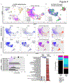
Figure 4.. YAP1 in LCNEC patient biopsies and PDX models is associated with higher EMT.
A, UMAP plot of all cell populations from 6 LCNEC and combined histology biopsies (left) and UMAP plot of cancer cell populations (right), both labeled by patient. B, UMAP plot of all cell populations from 6 LCNEC PDX models, labeled by model. C, UMAP plots demonstrating binary expression of ASCL1 and YAP1. YAP1-expression cell populations had elevated YAP/TAZ target scores and EMT scores. D, Percentage of YAP1-positive cells in each biopsy. E, GSEA of YAP1-positive and -negative cells. F. Subpopulation breakdown of cell populations in the YAP1-high and -low biopsies. There was a greater percentage of cancer cells in the YAP1-low biopsies (P<0.001) and a higher percentage of all immune cell populations in the YAP1-high biopsies (P<0.001).
Figure 5.
Figure 5.. YAP1-high LCNEC is sensitive to MEK1/2 inhibition.
YAP1 status does not predict sensitivity to cisplatin in LCNEC cell lines (A) or cell line/PDX xenograft models (B). C, Treatment of LCNEC cell lines with DMSO, cisplatin, MYF-01-37, XAV-939 and decitabine did not change YAP1, but verteporfin reduced YAP1 levels. D, YAP1 status does not predict response to verteporfin. E, YAP1-low cell lines had improved response to venetoclax (upper left). LCNEC cell lines were sensitive to obatoclax, alisertib, and 17-AAG with no difference in response based on YAP1 status. F, Waterfall plot of drug sensitivity in LCNEC cell lines. A comparison of high YAP1 levels and IC50 values identified several drugs with similar targets, including MEK1/2, CDK4/6, and Src family kinase inhibitors. G, Tumor growth curves from YAP1-high cell line xenografts H1915 (LCNEC) and SW1271 (SCLC) demonstrate a delay in tumor growth with trametinib treatment.
Figure 6.
Figure 6.. Cellular therapies targeting YAP1 subsets of LCNEC.
A, AXL is expressed by YAP1-high and DLL3/NCAM1 are expressed by YAP1-low patient tumors. B, Cell surface expression of AXL, DLL3 and CD56 in LCNEC cell lines by flow cytometry demonstrates a similar pattern. C, Cytotoxicity of NT-T, DLL3 CAR-T, CD56 CAR-T, and AXL CAR-Ts in subsets of LCNEC. D, Correlation between cell surface expression of AXL and cytotoxicity of AXL CAR-T in LCNEC cell lines. E, UMAP plots visualizing YAP1 (left) or AXL (red) and NCAM1 (blue) or AXL (red) and DLL3 (green) in 6 LCNEC patient biopsies. Very few cells express both DLL3 and AXL or NCAM1 and AXL, suggesting that distinct cell populations can be targeted via the surfaceome. F, Schematic demonstrating common features of YAP1-high and low subtypes of pulmonary LCNEC.

References

    1. Watson GA, Ahmed Y, Picardo S, Chew S, Cobbe S, Mahony C, et al. Unusual Sites of High-Grade Neuroendocrine Carcinomas: A Case Series and Review of the Literature. Am J Case Rep 2018;19:710–23 doi 10.12659/AJCR.908953. - DOI - PMC - PubMed
    1. Rekhtman N. Lung neuroendocrine neoplasms: recent progress and persistent challenges. Mod Pathol 2022;35(Suppl 1):36–50 doi 10.1038/s41379-021-00943-2. - DOI - PMC - PubMed
    1. Le Treut J, Sault MC, Lena H, Souquet PJ, Vergnenegre A, Le Caer H, et al. Multicentre phase II study of cisplatin-etoposide chemotherapy for advanced large-cell neuroendocrine lung carcinoma: the GFPC 0302 study. Ann Oncol 2013;24(6):1548–52 doi 10.1093/annonc/mdt009. - DOI - PubMed
    1. Huang L, Feng Y, Xie T, Zhu H, Tang L, Shi Y. Incidence, survival comparison, and novel prognostic evaluation approaches for stage iii-iv pulmonary large cell neuroendocrine carcinoma and small cell lung cancer. BMC Cancer 2023;23(1):312 doi 10.1186/s12885-023-10797-3. - DOI - PMC - PubMed
    1. Zhang JT, Li Y, Yan LX, Zhu ZF, Dong XR, Chu Q, et al. Disparity in clinical outcomes between pure and combined pulmonary large-cell neuroendocrine carcinoma: A multi-center retrospective study. Lung Cancer 2020;139:118–23 doi 10.1016/j.lungcan.2019.11.004. - DOI - PubMed

MeSH terms