. 2024 Dec;29(1):2392329.
doi: 10.1080/13510002.2024.2392329. Epub 2024 Aug 16.

Sarmentosin alleviates doxorubicin-induced cardiotoxicity and ferroptosis via the p62-Keap1-Nrf2 pathway

Affiliations

Sarmentosin alleviates doxorubicin-induced cardiotoxicity and ferroptosis via the p62-Keap1-Nrf2 pathway

Zhihui Lin et al. Redox Rep. 2024 Dec.

Abstract

Doxorubicin (Dox) is extensively used as an antitumor agent, but its severe cardiotoxicity significantly limits its clinical use. Current treatments for Dox-induced cardiotoxicity are inadequate, necessitating alternative solutions. This study evaluated the effects of sarmentosin, a compound from Sedum sarmentosum, on Dox-induced cardiotoxicity and dysfunction. Sarmentosin was administered as a pretreatment to both mice and H9c2 cells before Dox exposure. Subsequently, markers of Dox-induced cardiotoxicity and ferroptosis in serum and cell supernatants were measured. Western blot analysis was utilized to detect levels of ferroptosis, oxidative stress, and autophagy proteins. Additionally, echocardiography, hematoxylin-eosin staining, ROS detection, and immunofluorescence techniques were employed to support our findings. Results demonstrated that sarmentosin significantly inhibited iron accumulation, lipid peroxidation, and oxidative stress, thereby reducing Dox-induced ferroptosis and cardiotoxicity in C57BL/6 mice and H9c2 cells. The mechanism involved the activation of autophagy and the Nrf2 signaling pathway. These findings suggest that sarmentosin may prevent Dox-induced cardiotoxicity by mitigating ferroptosis. The study underscores the potential of compounds like sarmentosin in treating Dox-induced cardiotoxicity.

Keywords: Doxorubicin; Sarmentosin; autophagy; cardiotoxicity; ferroptosis.


Conflict of interest statement

No potential conflict of interest was reported by the author(s).

Figures
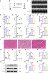
Figure 1.
Figure 1.
Cardiotoxicity and ferroptosis caused by Dox were alleviated by Sarmentosin in vivo. (A) Chemical structure of sarmentosin. (B) Establishment of the Dox model. (C) Representative echocardiogram images of each group. (D) LVEF, n = 6. (E) LVFS, n = 6. (F-H) Weight of the body, heart weight, and index of heart/tibia, n = 6. (I-K) Serum levels of LDH, CKMB, and cTnT in mice, n = 6. (L) The staining of HE in representative images (Scale bar: 50 µm), n = 6. (M-P) Myocardial iron content, MDA level, GSH level, and SOD level, n = 6. (Q) Assayed with Western blotting in vivo for PTGS2 and GPX4. (R-S) Expression of PTGS2 and GPX4 proteins quantitatively analyzed. A comparison was made between levels of normalized expression and levels of β-actin, n = 3. Data are means ± SD, *P < 0.05; ** P < 0.01.
Figure 2.
Figure 2.
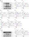
Cardiotoxicity and ferroptosis caused by Dox were alleviated by Sarmentosin in vitro. (A) Each group was tested for cell viability, n = 6. (B) In vitro levels of LDH, n = 6. (C-F) In H9c2 cells, the iron content, MDA level, GSH level, and SOD level were determined, n = 6. (G) C11-BODIPY fluorescence of lipid ROS in each group (Scale bar: 200 µm), n = 3. (H) Assayed with Western blotting in vitro for PTGS2 and GPX4. (I-J) Expression of PTGS2 and GPX4 proteins quantitatively analyzed. A comparison was made between levels of normalized expression and levels of β-actin, n = 3. Data are means ± SD, *P < 0.05; ** P < 0.01.
Figure 3.
Figure 3.
Sarmentosin adjusted Nrf2 signal pathway in vivo and in vitro. (A) Assayed with Western blotting in vivo for Keap1, Nrf2, HO-1 and NQO1. (B) Assayed with Western blotting in vitro for Keap1, Nrf2, HO-1 and NQO1. (C-F) Expression of Keap1, Nrf2, HO-1 and NQO1 proteins quantitatively analyzed in vivo. A comparison was made between levels of normalized expression and levels of β-actin n = 3. (G-J) Expression of Keap1, Nrf2, HO-1 and NQO1 proteins quantitatively analyzed in vitro. A comparison was made between levels of normalized expression and levels of β-actin, n = 3. (K) Immunofluorescence staining of Nrf2 in each group (Scale bar: 50 µm), n = 3. (L) Quantification of Nrf2 relative fluorescence intensity. Data are means ± SD, *P < 0.05; ** P < 0.01.
Figure 4.
Figure 4.
Sarmentosin alleviated doxorubicin-induced cardiotoxicity and ferroptosis via a Nrf2-dependent mechanism. (A) Assayed with Western blotting in vitro for Nrf2, HO-1 and NQO1. (B-D) Expression of Nrf2, HO-1 and NQO1 proteins quantitatively analyzed. A comparison was made between levels of normalized expression and levels of β-actin, n = 3. (E) Each group was tested for cell viability, n = 6. (F) In vitro levels of LDH, n = 6. (G-J) In H9c2 cells, the iron content, MDA level, GSH level, and SOD level were determined, n = 6. (K) Assayed with Western blotting in vitro for PTGS2 and GPX4. (L-M) Expression of PTGS2 and GPX4 proteins quantitatively analyzed. A comparison was made between levels of normalized expression and levels of β-actin, n = 3. Data are means ± SD, *P < 0.05; ** P < 0.01.
Figure 5.
Figure 5.
Sarmentosin treatment promoted autophagic flux in models of Dox-induced cardiotoxicity. (A) Assayed with Western blotting in vivo for Atg5, LC3B and P62. (B-D) Expression of Atg5, LC3B and P62 proteins quantitatively analyzed in vivo. A comparison was made between levels of normalized expression and levels of β-actin, n = 3. (E) Assayed with Western blotting in vitro for Atg5, LC3B and P62. (F-H) Expression of Atg5, LC3B and P62 proteins quantitatively analyzed in vitro. A comparison was made between levels of normalized expression and levels of β-actin, n = 3. (I) Immunofluorescence staining of LC3B in each group (Scale bar: 50 µm), n = 3. (J) Quantification of LC3B relative fluorescence intensity. (K) Representative images of H9c2 cells transfected Ad-mCherry-GFP-LC3B adenovirus in each group (Scale bar: 100 µm), n = 3. (L) Quantification of GFP-LC3B relative fluorescence intensity. Data are means ± SD, *P < 0.05; ** P < 0.01.
Figure 6.
Figure 6.
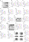
Sarmentosin ameliorated Dox-induced injury and inhibited ferroptosis by promoting autophagosome degradation in vitro. (A) Assayed with Western blotting in vivo for Atg5, P62 and LC3B. (B-D) Quantitative analysis of the expression of Atg5, P62 and LC3B proteins. A comparison was made between levels of normalized expression and levels of β-actin, n = 3. (E) Each group was tested for cell viability, n = 6. (F) In vitro levels of LDH, n = 6. (G-J) In H9c2 cells, the iron content, MDA level, GSH level, and SOD level were determined, n = 6 (K) Assayed with Western blotting in vitro for PTGS2 and GPX4. (L-M) Quantitative analysis of the expression of PTGS2 and GPX4 proteins. A comparison was made between levels of normalized expression and levels of β-actin, n = 3. (N) Assayed with Western blotting in vitro for Keap1, Nrf2, HO-1 and NQO1. (O-R) Quantitative analysis of the expression of Keap1, Nrf2, HO-1 and NQO1 proteins. A comparison was made between levels of normalized expression and levels of β-actin, n = 3. (S) Combination of Keap1 protein with P62 was identified by Co-IP. Normalized expression levels were compared to β-actin, n = 3. (T-U) Western blotting analysis of Co-IP, n = 3. Data are means ± SD, *P < 0.05; ** P < 0.01.
Figure 7.
Figure 7.
Schematic: As a result of its ability to promote autophagy and Nrf2 signaling, sarmamentosin relieves the cardiotoxicity and ferroptosis induced by doxorubicin.

References

    1. Perou CM, Sørlie T, Eisen MB, et al. . Molecular portraits of human breast tumours. Nature. 2000;406(6797):747–752. doi:10.1038/35021093 - DOI - PubMed
    1. Sørlie T, Perou CM, Tibshirani R, et al. . Gene expression patterns of breast carcinomas distinguish tumor subclasses with clinical implications. Proc Natl Acad Sci U S A. 2001;98(19):10869–10874. doi:10.1073/pnas.191367098 - DOI - PMC - PubMed
    1. Ji Y, Jin D, Qi J, et al. . Fucoidan protects against doxorubicin-induced cardiotoxicity by reducing oxidative stress and preventing mitochondrial function injury. Int J Mol Sci. 2022;23(18):10685. - PMC - PubMed
    1. Chen R, Sun G, Xu L, et al. . Didymin attenuates doxorubicin-induced cardiotoxicity by inhibiting oxidative stress. Chin Herb Med. 2022;14(1):70–78. - PMC - PubMed
    1. Peng Y, Wang L, Zhang Z, et al. . Puerarin activates adaptive autophagy and protects the myocardium against doxorubicin-induced cardiotoxicity via the 14-3-3γ/PKCϵ pathway. Biomed Pharmacother. 2022;153:113403. doi:10.1016/j.biopha.2022.113403 - DOI - PubMed

LinkOut - more resources