. 2024 Nov 14;45(43):4582-4597.
doi: 10.1093/eurheartj/ehae482.

The humanized platelet glycoprotein VI Fab inhibitor EMA601 protects from arterial thrombosis and ischaemic stroke in mice

Affiliations

The humanized platelet glycoprotein VI Fab inhibitor EMA601 protects from arterial thrombosis and ischaemic stroke in mice

Stefano Navarro et al. Eur Heart J. .

Abstract

Background and aims: Glycoprotein VI (GPVI) is a platelet collagen/fibrin(ogen) receptor and an emerging pharmacological target for the treatment of thrombotic and thrombo-inflammatory diseases, notably ischaemic stroke. A first anti-human GPVI (hGPVI) antibody Fab-fragment (ACT017/glenzocimab, KD: 4.1 nM) recently passed a clinical phase 1b/2a study in patients with acute ischaemic stroke and was found to be well tolerated, safe, and potentially beneficial. In this study, a novel humanized anti-GPVI antibody Fab-fragment (EMA601; KD: 0.195 nM) was developed that inhibits hGPVI function with very high potency in vitro and in vivo.

Methods: Fab-fragments of the mouse anti-hGPVI IgG Emf6.1 were tested for functional GPVI inhibition in human platelets and in hGPVI expressing (hGP6tg/tg) mouse platelets. The in vivo effect of Emf6.1Fab was assessed in a tail bleeding assay, an arterial thrombosis model and the transient middle cerebral artery occlusion (tMCAO) model of ischaemic stroke. Using complementary-determining region grafting, a humanized version of Emf6.1Fab (EMA601) was generated. Emf6.1Fab/EMA601 interaction with hGPVI was mapped in array format and kinetics and quantified by bio-layer interferometry.

Results: Emf6.1Fab (KD: 0.427 nM) blocked GPVI function in human and hGP6tg/tg mouse platelets in multiple assays in vitro at concentrations ≥5 µg/mL. Emf6.1Fab (4 mg/kg)-treated hGP6tg/tg mice showed potent hGPVI inhibition ex vivo and were profoundly protected from arterial thrombosis as well as from cerebral infarct growth after tMCAO, whereas tail-bleeding times remained unaffected. Emf6.1Fab binds to a so far undescribed membrane proximal epitope in GPVI. The humanized variant EMA601 displayed further increased affinity for hGPVI (KD: 0.195 nM) and fully inhibited the receptor at 0.5 µg/mL, corresponding to a >50-fold potency compared with ACT017.

Conclusions: EMA601 is a conceptually novel and promising anti-platelet agent to efficiently prevent or treat arterial thrombosis and thrombo-inflammatory pathologies in humans at risk.

Keywords: Anti-platelet therapy; GPVI; Platelet; Stroke; Thrombosis.


Figures

Structured Graphical Abstract
The novel humanized anti-glycoprotein VI (GPVI)-Fab, EMA601, very potently inhibits human GPVI signalling without blocking the ligand-binding site of the receptor. In vivo, EMA601 protects from arterial thrombosis and ischaemic stroke, while leaving tail bleeding times unaffected.
Figure 1
Figure 1
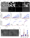
Emf6.1Fab inhibits glycoprotein VI–induced aggregation, thrombus formation, and spreading of human platelets. (A–E) Citrated blood from healthy donors was incubated with 5 µg/mL control Fab or Emf6.1Fab and then perfused over a collagen and tissue factor–coated surface (shear: 1000s−1). (A) Representative images; scale bar 50 µm. Platelet adhesion (B), thrombus formation (C), phosphatidylserine exposure (D), and fibrin/fibrinogen deposition (E) were assessed. Collagen and tissue factor were coated at 50 µg/mL and 500 pM, respectively. A flow rate of 1000 s−1 was used. Values are mean ± standard deviation (n = 4). A5: Annexin V, unpaired Mann–Whitney U test. **P < .01, ***P < .001. (F) Aggregation responses to the indicated agonists of washed human platelets pre-treated with 10 µg/mL Emf6.1Fab or control Fab in light-transmission aggregometry (n = 4). (G and H) Washed human platelets were allowed to spread on fibrinogen (100 µg/mL) for 45 min at 37°C. DIC pictures were taken (100× objective; scale bar 30 µm) (G) and phase abundance was determined. Phase 1: adhesion; Phase 2: filopodia formation; Phase 3: lamellipodia formation; Phase 4: fully spread platelet (H). Values are mean ± standard deviation (n = 8); unpaired, Mann–Whitney U test. ***P < .001
Figure 2
Figure 2
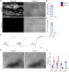
Emf6.1Fab inhibits glycoprotein VI–mediated aggregation, thrombus formation, and spreading of hGP6tg/tg platelets. (A–C) Assessment of platelet adhesion (B) and aggregate formation (C) on Horm collagen (200 µg/mL) under flow (1000 s−1) in heparinized hGP6tg/tg blood treated with 2 µg/mL of either Emf6.1Fab or control Fab. (A) Representative images are shown, scale 50 µm. Unpaired Mann–Whitney U test; values are mean ± standard deviation (n = 5); **P < .01, ***P < .001. (D) Aggregation responses of washed hGP6tg/tg platelets treated with either 5 µg/mL Emf6.1Fab or control Fab in light-transmission aggregometry (n = 5). Thr: Thrombin. (E and F) Washed hGP6tg/tg platelets were allowed to spread on fibrinogen (100 µg/mL) for 45 min at 37°C. DIC pictures were taken (100× objective, scale 30 µm) (E) and phase abundance was determined. Phase 1: adhesion; Phase 2: filopodia formation; Phase 3: lamellipodia formation; Phase 4: fully spread platelet (F). Values are mean ± standard deviation (n = 3); unpaired, Mann–Whitney U test.*P < .05, **P < .01
Figure 3
Figure 3
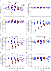
Emf6.1Fab efficiently blocks glycoprotein VI function in hGP6tg/tg mice. hGP6tg/tg animals (n = 5) were treated with 4 mg/kg b.w. Emf6.1Fab or control Fab. (A and B) Platelet count (A) and size (B) were determined using an automated cell counter. (C and D) Glycoprotein VI exposure was tested using Emf2 IgG-FITC (C), whereas epitope saturation of Emf6.1 was tested using Emf3 IgG-FITC (D). (E–H) Degranulation (α-P-selectin-FITC) (E and F) and activation of platelet αIIbβ3 integrin (JON/A-PE) (G and H) in 4 mg/kg b.w. Emf6.1Fab or control Fab-treated hGP6tg/tg mice was determined in flow cytometry upon activation with the indicated agonists. Data are expressed as mean ± standard deviation; significance is expressed as *P < .05, ***P < .001, vs. indicated group (two-way analysis of variance followed by Bonferroni’s multiple comparison test; n = 5)
Figure 4
Figure 4
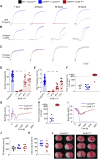
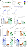
Emf6.1Fab treatment (4 mg/kg b.w.) protects mice from occlusive arterial thrombosis and ischaemic stroke without impairing haemostasis. (A–C) Aggregation responses of washed platelets from hGP6tg/tg mice intravenously treated with 4 mg/kg b.w. Emf6.1Fab or control Fab to collagen-related peptide (A), collagen (B), or thrombin (C) in light-transmission aggregometry (n = 6). (D and E) Assessment of platelet adhesion (D) and aggregate formation (E) on Horm collagen (200 µg/mL) under flow (1000 s−1) in heparinized blood from hGP6tg/tg mice intravenously treated with 4 mg/kg b.w. Emf6.1Fab or control Fab solution at different time points after injection. Values are mean ± standard deviation. Two-way analysis of variance followed by Bonferroni’s multiple comparison test *P < .05, ***P < .001. (F and G). Arterial thrombosis was assessed for a maximum period of 30 min after control Fab or Emf6.1Fab treatment in the mechanical injury of the aorta thrombosis model. (F) Time to vessel occlusion is depicted with each symbol representing one animal. ***P < .001 using Fisher’s exact test. (G) Representative blood flow traces are shown from control Fab and Emf6.1Fab-treated mice. (H and I) Arterial thrombosis was assessed for a maximum period of 30 min after control Fab or Emf6.1Fab treatment by topically injuring the carotid artery with a saturated filter paper with 10% FeCl3 for 3 min and time to occlusion was determined (H). Each symbol represents one individual. Representative blood flow traces are shown from control Fab and Emf6.1Fab-treated mice (I). ***P < .001 using Fisher’s exact test. (J) Haemostasis was assessed using the tail bleeding time with each symbol representing one mouse. (K and L) Cerebral infarct development after experimental stroke. (K) Oedema-corrected quantifications of infarct sizes from mice either treated with 4 mg/kg Emf6.1Fab or control before transient middle cerebral artery occlusion. (L) Representative images of brain sections stained with triphenyltetrazolium chloride to visualize infarcts. Each column of images represents three consecutive sections of one mouse. Data are depicted as mean ± standard deviation. Significance is expressed as *P < .05 using the Mann–Whitney U test
Figure 5
Figure 5
Emf6.1 binds a novel glycoprotein VI epitope. Glycoprotein VI-epitope mapping and confirmation of Emf6.1. (A) 15-Mer overlapping peptides covering the ectodomain (Residues 24–266) of glycoprotein VI were displayed in peptide-microarray format. Emf6.1 IgG binding was visualized by HRP-labelled anti-mouse secondary antibody. Mapping identified two distinct epitopes 201V-P215 and 246S-P260. (B) Emf6.1 binding is neutralized in the presence of the soluble glycoprotein VI peptide fragments 201V-P215 and 246S-P260 or vehicle solution. (C) Surface representation of glycoprotein VI (PDB-ID 2gi7). The Emf6.1-binding epitope (red) overlaps with the putative dimerization site (green) of glycoprotein VI but not the substrate-binding site (shown in orange for collagen PDB-ID 5ou9). The PyMOL image was generated by aligning the AF2 prediction (generated with AlphaFold) to glycoprotein VI–collagen (5ou9) and the dimer (2gi7). Data are expressed as mean ± standard deviation; significance is expressed as *P < .05
Figure 6
Figure 6
EMA601 inhibits glycoprotein VI function. (A–C) Assessment of platelet adhesion (B) and aggregate formation (C) on Horm collagen (50 µg/mL) under flow (1000 s−1) in heparinized hGP6tg/tg blood treated with 5 µg/mL EMA601 or control Fab. Values are mean ± standard deviation (n = 3). Unpaired, Mann–Whitney U test *P < .05, **P < .01. (A) Representative images are shown, scale 50 µM. (D) Aggregation responses of washed hGP6tg/tg platelets treated with either 5 µg/mL EMA601 or control Fab in light-transmission aggregometry (n = 3). (E and F) Washed hGP6tg/tg platelets were allowed to spread on fibrinogen (100 µg/mL) for 45 min at 37°C. DIC pictures were taken (100× objective, scale 30 µM) (E) and phase abundance was determined. Phase 1: adhesion; Phase 2: filopodia formation; Phase 3: lamellipodia formation; Phase 4: fully spread platelet (F). Values are mean ± standard deviation (n = 3); unpaired, Mann–Whitney U test *P < .05, **P < .01. (G–K) Assessment of platelet adhesion (H), thrombus formation (I), phosphatidylserine exposure (J), and fibrin/fibrinogen deposition (K) on Horm collagen (200 µg/mL) plus tissue factor (500 pM) under flow (1000 s−1) in recalcified human blood treated with 5 µg/mL EMA601 or control Fab. Values are mean ± standard deviation (n = 4). Unpaired, Mann–Whitney U test. ***P < .001. (D) Representative images are shown, scale 50 µm
Figure 7
Figure 7
EMA601 inhibits glycoprotein VI function with >50-fold potency compared with ACT017. hGP6tg/tg diluted blood (n = 4) was pre-incubated with the indicated concentrations (µg/mL) of either EMA601 or ACT017 and interference with Emf3FITC binding (A) was detected by flow cytometry. (B) Diluted blood of hGP6tg/tg (n = 4) mice was pre-incubated with the indicated concentrations of EMA601 or ACT017 and binding to platelets was detected using an anti-human IgG (Fab specific) antibody. (C and D) Aggregation traces of washed human platelets pre-treated with the indicated concentrations of EMA601 or ACT017 and stimulated with collagen (C) or collagen-related peptide (D). (E and F) Assessment of platelet adhesion (E) and aggregate formation (F) on Horm collagen (200 μg/mL) under flow (1000 s−1) in heparinized human blood treated with the indicated concentrations of EMA601, ACT017, or control Fab. (G–J) hGP6tg/tg animals were treated with 4 mg/kg (G and I) or 10 mg/kg (H and J) b.w. EMA601, ACT017, or control Fab. At the indicated time points after treatment, epitope saturation was tested using the anti-human IgG (Fab specific) antibody (G and H), while the activation of platelet αIIbβ3 integrin in response to 0.5 µg/mL collagen-related peptide was measured using JON/A-PE (I and J). Data are expressed as mean ± standard deviation; significance is expressed as *P < .05, **P < .01, ***P < .001, vs. indicated group (two-way analysis of variance followed by Bonferroni’s multiple comparison test)

Similar articles

Cited by

References

    1. Michelson AD, Cattaneo M, Frelinger A, Newman P. Platelets. 4th edn. Amsterdam, The Netherlands: Elsevier; 2019.
    1. Jackson SP. Arterial thrombosis–insidious, unpredictable and deadly. Nat Med 2011;17:1423–36. 10.1038/nm.2515 - DOI - PubMed
    1. Stellos K, Bigalke B, Borst O, Pfaff F, Elskamp A, Sachsenmaier S, et al. . Circulating platelet-progenitor cell coaggregate formation is increased in patients with acute coronary syndromes and augments recruitment of CD34+ cells in the ischaemic microcirculation. Eur Heart J 2013;34:2548–56. 10.1093/eurheartj/eht131 - DOI - PubMed
    1. Rodriguez F, Harrington RA. Management of antithrombotic therapy after acute coronary syndromes. N Engl J Med 2021;384:452–60. 10.1056/NEJMra1607714 - DOI - PMC - PubMed
    1. Stoll G, Nieswandt B. Thrombo-inflammation in acute ischaemic stroke—implications for treatment. Nat Rev Neurol 2019;15:473–81. 10.1038/s41582-019-0221-1 - DOI - PubMed

MeSH terms

Substances