. 2024 Aug 16;10(33):eadk0015.
doi: 10.1126/sciadv.adk0015. Epub 2024 Aug 16.

A high-throughput microfabricated platform for rapid quantification of metastatic potential

Affiliations

A high-throughput microfabricated platform for rapid quantification of metastatic potential

Smiti Bhattacharya et al. Sci Adv. .

Abstract

Assays that measure morphology, proliferation, motility, deformability, and migration are used to study the invasiveness of cancer cells. However, native invasive potential of cells may be hidden from these contextual metrics because they depend on culture conditions. We created a micropatterned chip that mimics the native environmental conditions, quantifies the invasive potential of tumor cells, and improves our understanding of the malignancy signatures. Unlike conventional assays, which rely on indirect measurements of metastatic potential, our method uses three-dimensional microchannels to measure the basal native invasiveness without chemoattractants or microfluidics. No change in cell death or proliferation is observed on our chips. Using six cancer cell lines, we show that our system is more sensitive than other motility-based assays, measures of nuclear deformability, or cell morphometrics. In addition to quantifying metastatic potential, our platform can distinguish between motility and invasiveness, help study molecular mechanisms of invasion, and screen for targeted therapeutics.


Figures

Fig. 1.
Fig. 1.. Microfabricated platform on glass allows tracking of cells with high spatial resolution during basal invasion activity.
(A) Schematic representation of the photolithographic process used to fabricate the platform. UV, ultraviolet. A negative-tone SU-8-like photoresist was used to transfer (B) a repeated array of 2500-μm2 square chambers interconnected with 5-μm-wide (width) and 30-μm-long (length) channels. (C) Scanning electron microscopy (SEM) of 3D micropatterns shows cellular compliance within the patterns. (D) Schematic showing how a micropatterned substrate is integrated with standard 96-well plates allowing for controlled multiplexed experiments with different cell types or antimetastatic drug candidates. (E) Representative immunofluorescent images of triple-negative LM2-4 and MCF-7 breast cancer cells plated on micropatterns representing cellular distribution 1 day after seeding on the micropatterns. (F) Still frame high-resolution image sequence showing a triple-negative LM2-4 cell (green) squeezing through the length of a channel (purple) as imaged in 3D on a modified lattice light-sheet microscope.
Fig. 2.
Fig. 2.. Microchannel assay can be used to quantify metastatic potential.
(A) Mapped trajectory of the nucleus of an MCF-7 (pseudocolored blue) and (B) triple-negative LM2-4 breast cancer cell line (pseudocolored red) seeded on micropatterns. Nuclei tagged with NucBlue were imaged over 24 hours. Captured images were subsequently superimposed to show cell trajectories. Less invasive MCF-7 cells remain trapped within the 2500-μm2 chambers, whereas highly metastatic LM2-4 cells invade into neighboring chambers through the narrow channels by squeezing their nuclei. (C) The mean total distance traveled by the migrating LM2-4 cells (n = 337) is significantly higher than that of MCF-7 cells (n = 252). (D) Cell invasion is quantified by computing the ratio of cellular invasions crossing in between interconnected chambers to the total number of cells visible in a viewing area. Invasion index for MCF-7 and LM2-4 breast cancer cells in the micropatterns shows nearly two orders of magnitude difference between the metastatic line compared to the nonmetastatic line. (E) When computed, the ratio of the invasion index to the distance traveled is roughly two orders of magnitude higher for the LM2-4 reiterating the high metastatic potential of this cell line. (F) A normalized histogram of the number of crossing events of the two cell types shows the percentage of cells in the viewing area that have a greater tendency to “invade” (**P < 0.001 and ****P < 0.0001).
Fig. 3.
Fig. 3.. Microchannel assay is more sensitive than current state-of-the-art high-throughput platforms.
(A) Crystal violet–stained representative images of MCF-7 and LM2-4 cells migrating in the Transwell chamber migration assay, under the same conditions as those of the micropattern assay, confirm that the inferred migration of LM2-4 cells is significantly higher than that of MCF-7 cells (scale bars, 20 μm). (B) Similarly, representative bright-field microscopy images of MCF-7 and LM2-4 cells migrating in a 2D assay, where a confluent monolayer is “injured” using a 0.5-mm-wide pipette tip, under the same experimental conditions as those of the micropattern assay, show significantly faster wound closure in LM2-4 cells within a 24-hour window (scale bars, 200 μm; ****P < 0.0001). (C) The effective sensitivities of the three assays, as computed by taking the ratio of inferred invasion of the metastatic LM2-4 cell line versus that of the nonmetastatic MCF-7 cell line, show that the microchannel assay exhibits superior sensitivity.
Fig. 4.
Fig. 4.. Decreased nuclear stiffness correlates with invasiveness, but it is not the principal factor.
(A) Representative phase contrast images of the six cell lines probed by AFM elastography. (B) The six tumor cell lines [MCF-7 (breast, n = 54), Huh7 (liver, n = 56), HepG2 (liver, n = 60), MDA-MB-468 (breast, n = 51), LM2-4 (breast, n = 56), and LCC6 (breast, n = 21)] were probed around their nuclear regions via AFM indentations. Nuclear stiffness of both LM2-4 and LCC6 cell lines was significantly lower compared to all the other cell lines (see table S1). This was coupled with an increase in the metastatic potential as seen in both the (C) microchannel and (D) Transwell assays run for all six cell types. Both LM2-4 and LCC6 cell lines had significantly higher invasion indices compared to the other cell lines (see table S2). This trend was similar in the average Transwell absorbance as well (see table S3). *P < 0.01.
Fig. 5.
Fig. 5.. Microchannel assay can separate effects on invasiveness and motility in drug screening.
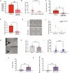
Results from representative antimetastatic drug efficacy experiments, where LM2-4 cells cultured with charcoal-stripped medium (LM2-4cs) were treated by either vehicle or a small-molecule TKI, BMS-754807, that inhibits IR/IGFR1 activity. (A) Percent wound closure in the scratch assay for LM2-4 cells versus those treated with BMS-754807. (B) TKI-treated LM2-4 cells plated on the microchannel assay showed a significant decrease in total distance traveled. (C) The invasion index of TKI-treated LM2-4 cells also showed a significant decrease compared to its untreated counterpart. (D) This trend remained when invasion index was normalized invasion to the total displacement. (E) To unambiguously separate motility from invasion, we created an open layout version of our micropatterned surfaces, where cells can migrate freely. (F) Normalized amount of intra-chamber motion (i.e., invasion index for the 5-μm layout) showed a decrease in both versions though the interaction term was not statistically significant per two-way analysis of variance (ANOVA) with Bonferroni correction. (G) Representative phase contrast images of vehicle and trichostatin A (TSA)–treated MCF-7 cells probed by AFM elastography. (H) The MCF-7 cell line at baseline (n = 22) and after TSA treatment (n = 21), when probed around the nuclear region, showed a significant decrease in stiffness. (I) The total distance traveled by MCF-7 cells treated with TSA (n = 462) exhibits a significant decrease when compared with the untreated MCF-7 cells (n = 776). (J) The corresponding invasion index shows a significant increase in the treated versus control cells, an increase that is much higher for (K) the invasion index normalized to the total traveled distance (*P < 0.01, **P < 0.001, and ****P < 0.0001).

References

    1. Chaffer C. L., Weinberg R. A., A perspective on cancer cell metastasis. Science 331, 1559–1564 (2011). - PubMed
    1. Kortam S., Merkher Y., Kramer A., Metanes I., Ad-el D., Krausz J., Har-Shai Y., Weihs D., Rapid, quantitative prediction of tumor invasiveness in non-melanoma skin cancers using mechanobiology-based assay. Biomech. Model. Mechanobiol. 20, 1767–1774 (2021). - PubMed
    1. Merkher Y., Horesh Y., Abramov Z., Shleifer G., Ben-Ishay O., Kluger Y., Weihs D., Rapid cancer diagnosis and early prognosis of metastatic risk based on mechanical invasiveness of sampled cells. Ann. Biomed. Eng. 48, 2846–2858 (2020). - PubMed
    1. Wullkopf L., West A. K. V., Leijnse N., Cox T. R., Madsen C. D., Oddershede L. B., Erler J. T., Cancer cells’ ability to mechanically adjust to extracellular matrix stiffness correlates with their invasive potential. Mol. Biol. Cell 29, 2378–2385 (2018). - PMC - PubMed
    1. Hu J., Zhou Y., Obayemi J. D., Du J., Soboyejo W. O., An investigation of the viscoelastic properties and the actin cytoskeletal structure of triple negative breast cancer cells. J. Mech. Behav. Biomed. Mater. 86, 1–13 (2018). - PubMed

Publication types

LinkOut - more resources