. 2024 Aug 16;10(33):eado1533.
doi: 10.1126/sciadv.ado1533. Epub 2024 Aug 16.

Epinephrine promotes breast cancer metastasis through a ubiquitin-specific peptidase 22-mediated lipolysis circuit

Affiliations

Epinephrine promotes breast cancer metastasis through a ubiquitin-specific peptidase 22-mediated lipolysis circuit

Yuanzhang Zhou et al. Sci Adv. .

Abstract

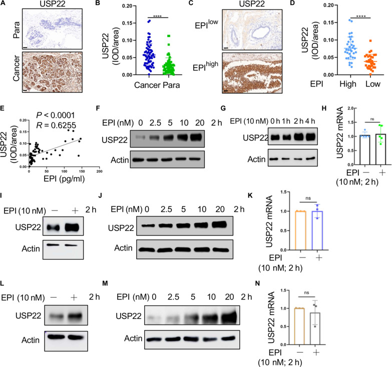
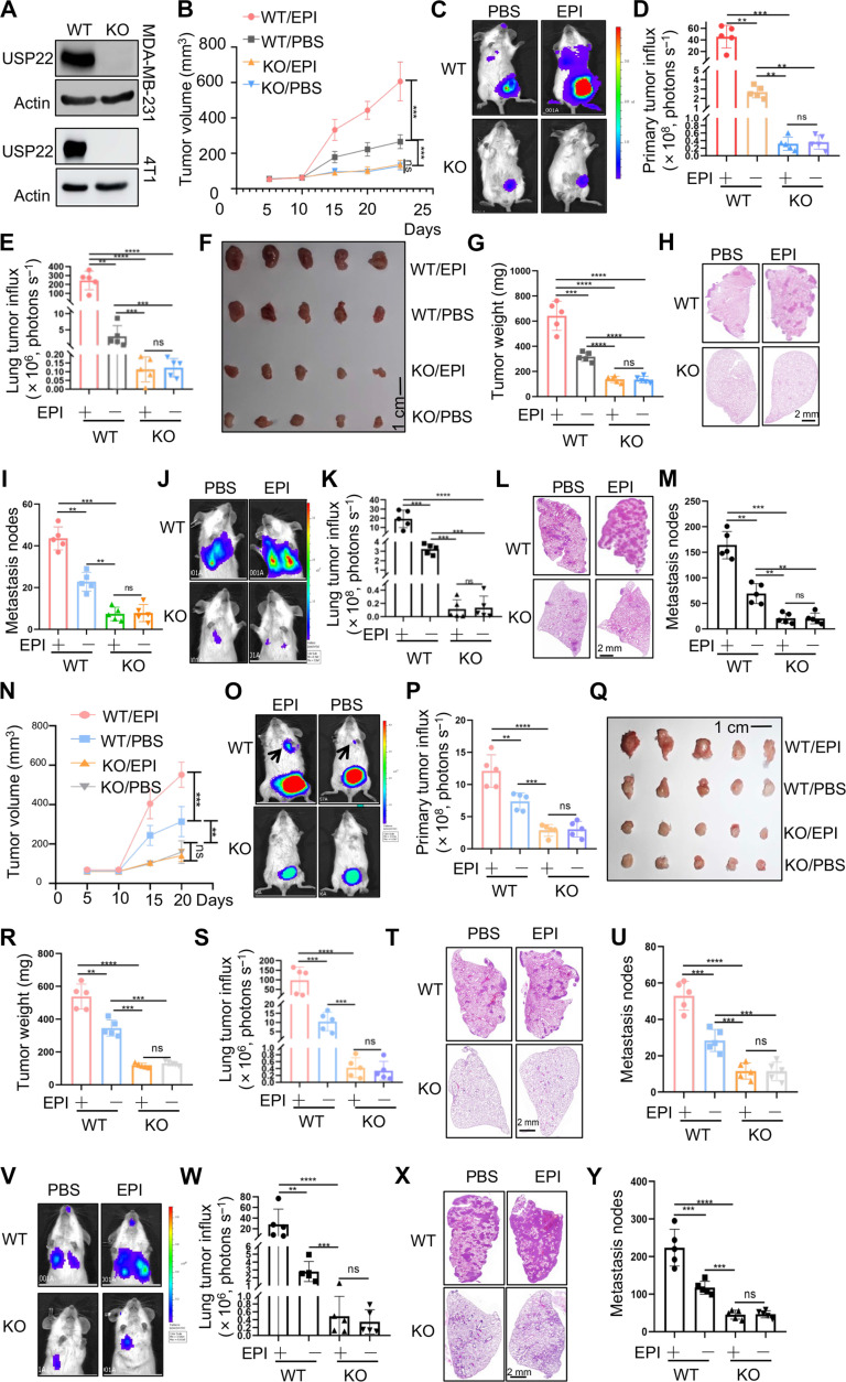
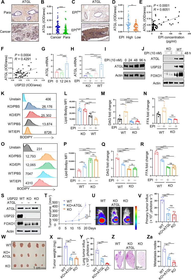
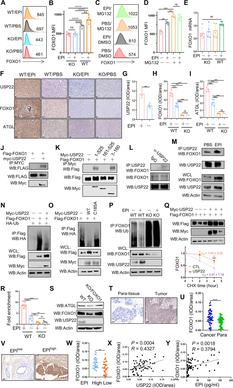
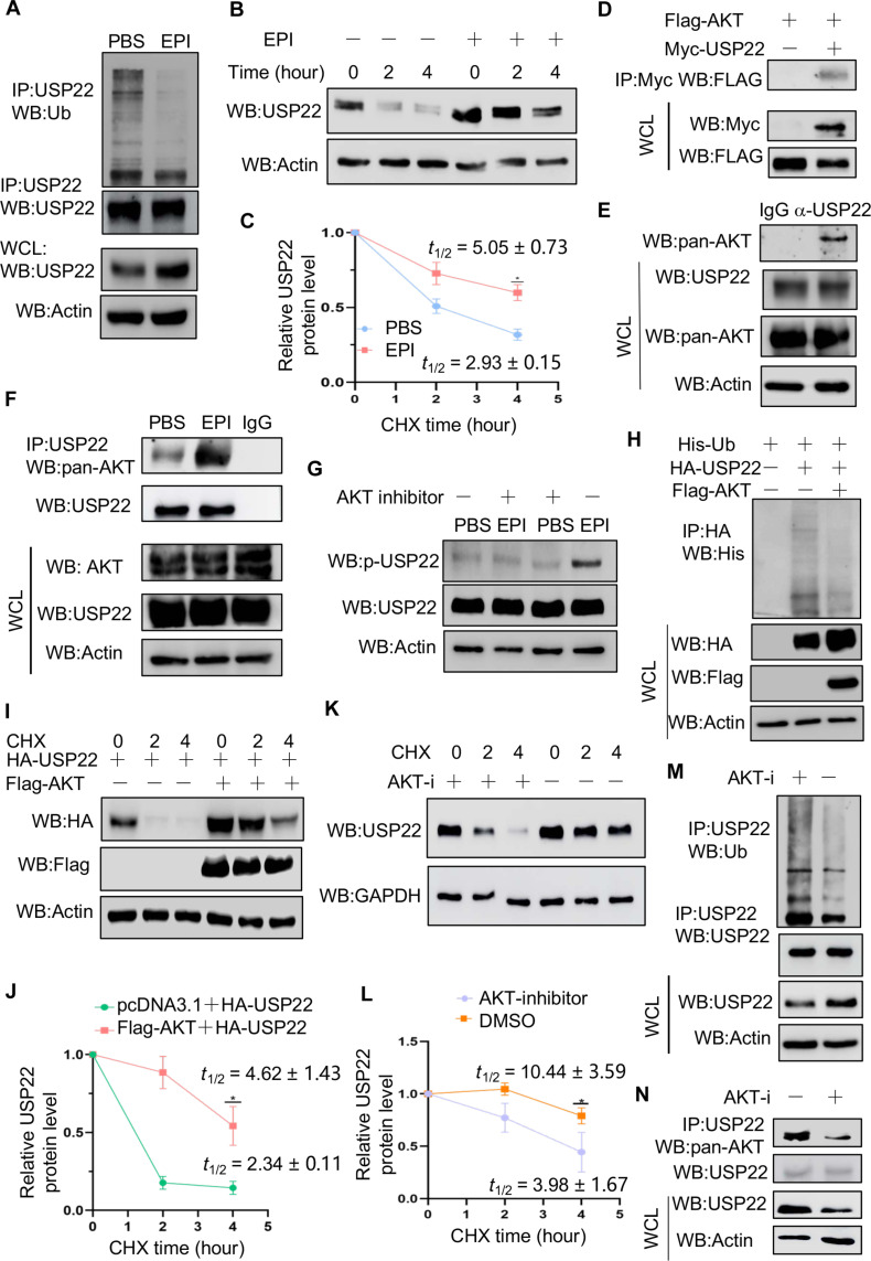
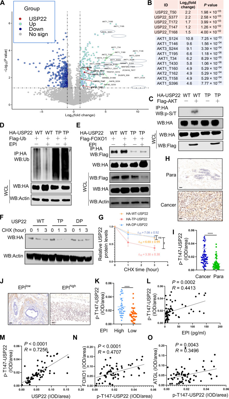
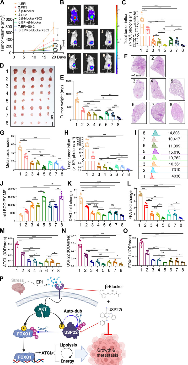
Chronic stress-induced epinephrine (EPI) accelerates breast cancer progression and metastasis, but the molecular mechanisms remain unclear. Herein, we found a strong positive correlation between circulating EPI levels and the tumoral expression of ubiquitin-specific peptidase 22 (USP22) in patients with breast cancer. USP22 facilitated EPI-induced breast cancer progression and metastasis by enhancing adipose triglyceride lipase (ATGL)-mediated lipolysis. Targeted USP22 deletion decreased ATGL expression and lipolysis, subsequently inhibiting EPI-mediated breast cancer lung metastasis. USP22 acts as a bona fide deubiquitinase for the Atgl gene transcription factor FOXO1, and EPI architects a lipolysis signaling pathway to stabilize USP22 through AKT-mediated phosphorylation. Notably, USP22 phosphorylation levels are positively associated with EPI and with downstream pathways involving both FOXO1 and ATGL in breast cancers. Pharmacological USP22 inhibition synergized with β-blockers in treating preclinical xenograft breast cancer models. This study reveals a molecular pathway behind EPI's tumor-promoting effects and provides a strong rationale for combining USP22 inhibition with β-blockers to treat aggressive breast cancer.


Figures

Fig. 1.
Fig. 1.. High EPI in patients with breast cancer is accompanied with the increasing expression of USP22.
(A) Representative IHC photomicrographs of tissues stained with USP22 antibodies in patients with breast cancer in clinical breast cancer tissues and para-cancer tissues; Scale bars, 50 μm. (B) Quantification of USP22 expression in breast cancer tissues and para-cancer tissues (n = 65). (C and D) Representative images of USP22 staining in patients with breast cancer (EPIlow, n = 33; EPIhigh, n = 32); Scale bars, 50 μm (C). Analysis of USP22 expression in human breast cancer tissues in patients with high and low levels of EPI (D). (E) Relationship between serum EPI levels and USP22 from the IHC staining score in human breast cancer tissues. Pearson correlation coefficient was used as a measure of association. (F to N) Analysis of USP22 protein expression in MDA-MB-231 [(F) to (H)], MCF-7 [(I) to (K)], and SK-BR-3 [(L) to (N)] breast cancer cells treated with indicated concentrations of EPI [(F), (J), and (M)] or time course [(G), (I), and (L)]. Expression of mRNA of USP22 was measured by quantitative RT-PCR [(H), (K), and (N)]; Pearson correlation coefficient was used as a measure of association. Data are expressed as means ± SD of three or more independent experiments. Statistical significance was concluded by unpaired Student’s t test. ****P < 0.0001; ns, no statistical significance.
Fig. 2.
Fig. 2.. EPI increases the expression of ATGL to induce the lipolysis of breast cancer through USP22.
(A) Validation of USP22 protein expression in WT and USP22 KO MDA-MB-231 cells (top) or 4T1 cells (bottom). (B to I) A total of 1 × 10⁶ WT or USP22 KO MDA-MB-231 cells were injected into the mammary fat pad of immune compromised NYG mice and treated with PBS or EPI (2 mg/kg) per day for 7 days. Tumor volume (B), bioluminescence activity [(C) and (D)], and tumor weight [(F) and (G)] were measured. Lung metastases were measured by luminol fluorescence [(C) and (E)] and H&E staining of their lung sections [(H) and (I)]. N = 5 each group. (J to M) A total of 0.5 × 10⁶ WT or USP22 KO MDA-MB-231 cells were injected via the tail vein into NYG mice. Lung metastases were measured by luminol fluorescence [(J) and (K)] and H&E staining [(L) and (M)]. (N to U) A total of 1 × 10⁵ WT or USP22 KO 4T1 cells were injected into the mammary fat pad of BALB/c mice and treated with PBS or EPI (2 mg/kg) per day for 7 days. Tumor volume (N), bioluminescence activity [(O) and (P)], and tumor weight [(Q) and (R)] were measured. Lung metastases were measured by luminol fluorescence [(O) and (S)] and H&E staining of their lung sections [(T) and (U)]. N = 5 each group. (V to Y) A total of 0.5 × 10⁵ WT or USP22 KO 4T1 cells were injected via the tail vein into BALB/c mice. Lung metastases were measured by luminol fluorescence [(V) and (W)] and H&E staining [(X) and (Y)]. Statistical significance was determined by one-way ANOVA test or unpaired Student’s t test. Data are expressed as means ± SD of five independent experiments. **P < 0.01; ***P < 0.001; ****P < 0.0001; ns, no statistical significance.
Fig. 3.
Fig. 3.. USP22 promotes ATGL transcription and lipolysis in breast cancer cells.
(A to F) ATGL expression in breast cancer tissues by IHC staining. Representative images from para-tumor normal tissue and cancer sections (A) and quantification from 65 patients (B) are shown. Scale bars, 50 μm. The data were further analyzed by EPI levels [(C) and (D)]. The correlation of ATGL with EPI (E) and USP22 (F) was analyzed. (G to J) The mRNA [(G) and (H)] and protein [(I) and (J)] levels of ATGL in MDA-MB-231 cells treated with EPI (10 nM) were determined by real-time PCR and Western blotting, respectively. (K to M) Intracellular lipid analysis, including BODIPY [(K) and (L)], DAG (M), and FFAs (N) in WT and USP22-null MDA-MB-231 cells treated with EPI in vitro. (O to R) Intracellular lipid analysis in WT and USP22-null MDA-MB-231 cells isolated from the xenograft tumors as shown in Fig. 2E. (S) The protein levels of USP22, FOXO1, and ATGL in the WT 4T1 cells, USP22 KO 4T1 cells, and the USP22 KO 4T1 cells transduced with ATGL were measured. (T to Za) A total of 1 × 10⁵ WT, USP22 KO 4T1 cells, or USP22 KO 4T1 cells transduced with ATGL were injected into the mammary fat pad of BALB/c mice. Tumor volume (T), bioluminescence activity [(U) and (V)], and tumor weight [(W) and (X)] were measured. Lung metastases were measured by luminol fluorescence [(U) and (Y)] and H&E staining of their lung sections [(Z) and (Za)]. N = 5 each group. Pearson correlation coefficient was used as a measure of association. Statistical significance was determined by one-way ANOVA test or unpaired Student’s t test. Data are expressed as means ± SD of five independent experiments. *P < 0.05; **P < 0.01; ***P < 0.001; ns, no statistical significance.
Fig. 4.
Fig. 4.. USP22 is a deubiquitinase for Atgl transcription factor FOXO1.
(A to E) Effect of EPI on FOXO1 protein expression in WT and USP22-null MDA-MB-231 cells without [(A) and (B)] or with MG132 treatment [(C) and (D)]. The mRNA levels of FOXO1 were analyzed by real-time RT-PCR (E). (F to I) Analysis of USP22, FOXO1, and ATGL expression levels by IHC in syngeneic tumor tissues as shown in Fig. 2D. Representative images (F) (scale bars, 50 μm) and quantification data [(G) to (I)] are shown. (J to M) FOXO1 interaction in HEK239T cells transiently transfected with FOXO1 and USP22 (J) or USP22 truncated mutants (K) expressing plasmids and in MDA-MB-231 cells (L) or further with EPI treatment (M). WB, Western blotting. WCL, whole cell lysate. (N to P) Analysis of FOXO1 ubiquitination in HEK293T cells coexpressed with USP22 (N) and its mutants (O) and in USP22 WT and KO MDA-MB-231 cells (P). (Q) Immunoblot analysis of FOXO1 protein degradation in HEK293T cells transfected with or without USP22 (n = 3). (R) Analysis of FOXO1 binding to Atgl promoter by ChIP in WT and USP22 KO MDA-MB-231 cells (n = 5). (S) Reconstitution of FOXO1 expression in USP22-null MDA-MB-231 cells restored ATGL expression. (T to Y) Analysis of FOXO1 expression in human breast cancer tissues [(T) and (U) with para-tumor tissues as controls or (V) and (W) in EPIhigh and EPIlow groups] as well as its correlation with USP22 (X) and EPI (Y). Scale bars, 50 μm. Pearson correlation coefficient was used as a measure of association. Statistical significance was determined by one-way ANOVA test or unpaired Student’s t test. Data are expressed as means ± SD of three or more independent experiments. *P < 0.05; **P < 0.01; ***P < 0.001; ****P < 0.0001. ns, no statistical significance.
Fig. 5.
Fig. 5.. EPI promotes the phosphorylation of USP22 by AKT to enhance the stability of USP22.
(A to C) The effect of EPI on USP22 ubiquitination (A) and protein stability [(B) and (C)] in MDA-MB-231 cells were determined (n = 3). (D and E) Co-IP and immunoblot analysis of USP22 interaction with AKT in transiently transfected HEK293T cells (D) and MDA-MB-231 cells (E). (F and G) Effect of EPI on USP22 interaction with AKT and USP22 phosphorylation in MDA-MB-231 cells. (H to J) Effect of AKT overexpression on USP22 ubiquitination (H) and protein stability [(I) and (J)] (n = 3). (K to N) Effect of AKT inhibition on USP22 protein degradation [(K) and (L)] (n = 3), ubiquitination (M), and interaction with AKT (N) in MDA-MB-231 cells. AKT-i, AKT inhibitor. Statistical significance was determined by unpaired Student’s t test. Data are expressed as means ± SD of three independent experiments. *P < 0.05.
Fig. 6.
Fig. 6.. AKT-mediated USP22 phosphorylation controls its protein stability.
(A and B) Proteomic analysis of USP22 phosphorylation from HEK293T cells transfected with or without Flag-AKT. The spectra of peptides carrying phosphorylated peptides (A) and the identified phosphorylation residues of USP22 (top) and AKT (bottom) (B) are shown. (C and D) Analysis of phosphorylation (C) and ubiquitination (D) with USP22 and its phosphorylation mutant. (E) Analysis of FOXO1 interaction with USP22 and its phosphorylation mutant. (F and G) Analysis of protein stability of USP22 and its phosphorylation mutant (n = 3). (H to O) Analysis of USP22 phosphorylation in human breast cancer by IHC. Representative images in tumor and para-controls (H) or in EPIhigh and EPIlow groups (J) as well as the quantification from 65 patients [(I) and (K)] are shown. Scale bars, 50 μm. Statistical analyses of the correlation p-USP22-Thr147 levels with EPI (L), total USP22 (M), FOXO1 (N), and ATGL (O) are shown. Pearson correlation coefficient was used as a measure of association. Statistical significance was determined by unpaired Student’s t test. Data are expressed as means ± SD of three or more independent experiments. **P < 0.01; ****P < 0.0001; ns, no statistical significance.
Fig. 7.
Fig. 7.. Synergistic effects of USP22 and β receptor inhibitors on tumors.
(A to E) 4T1 TNBC cells were injected into the mammary fat pad of BALB/c mice (N = 5 each group). Tumor volume (A), bioluminescence activity [(B) and (C)], and tumor weight [(D) and (E)] were measured. (F to H) The metastases of the lung were measured by luminol fluorescence [(B) and (H)] and H&E staining [(F) and (G)]. Lung tissue sections were analyzed by H&E staining [(F) and (G)]. Representative lung metastasis (F) and quantifications of five mice each group (G) are shown. (I to L) Lipid analysis of syngeneic tumors from (D) by lipid BODIPY [(I) and (J)], DAG (K), and FFAs (L). MFI, mean fluorescence intensity. (M to O) IHC analysis of USP22, FOXO1, and ATGL expression levels in syngeneic tumor tissue sections from (D). Quantification data of all five mice each group [(M) to (O)] are shown. (P) Model of targeting chronic stress–mediated cancer metastasis and lipolysis by USP22. Statistical significance was determined by one-way ANOVA test or unpaired Student’s t test. Data are expressed as means ± SD of five independent experiments. *P < 0.05; **P < 0.01; ***P < 0.001; ****P < 0.0001; ns, no statistical significance.

References

    1. Weigelt B., Peterse J. L., van’t Veer L. J., Breast cancer metastasis: Markers and models. Nat. Rev. Cancer 5, 591–602 (2005). - PubMed
    1. Spiegel D., Bloom J. R., Kraemer H. C., Gottheil E., Effect of psychosocial treatment on survival of patients with metastatic breast cancer. Lancet 2, 888–891 (1989). - PubMed
    1. Borgi M., Collacchi B., Ortona E., Cirulli F., Stress and coping in women with breast cancer: Unravelling the mechanisms to improve resilience. Neurosci. Biobehav. Rev. 119, 406–421 (2020). - PubMed
    1. Zhu Z., Yu R., Yang C., Li D., Wang J., Yang W., Ji Y., Wang L., Wang Y., Jiang F., Stress-related hormone reduces autophagy through the regulation of phosphatidylethanolamine in breast cancer cells. Ann. Transl. Med. 9, 149 (2021). - PMC - PubMed
    1. Chang A., Botteri E., Gillis R. D., Lofling L., Le C. P., Ziegler A. I., Chung N. C., Rowe M. C., Fabb S. A., Hartley B. J., Nowell C. J., Kurozumi S., Gandini S., Munzone E., Montagna E., Eikelis N., Phillips S. E., Honda C., Masuda K., Katayama A., Oyama T., Cole S. W., Lambert G. W., Walker A. K., Sloan E. K., Beta-blockade enhances anthracycline control of metastasis in triple-negative breast cancer. Sci. Transl. Med. 15, eadf1147 (2023). - PubMed

Publication types

MeSH terms