. 2024 Sep 10;103(5):e209770.
doi: 10.1212/WNL.0000000000209770. Epub 2024 Aug 16.

Temporal and Spatial Clustering of Intracerebral Hemorrhage in Cerebral Amyloid Angiopathy

Affiliations

Temporal and Spatial Clustering of Intracerebral Hemorrhage in Cerebral Amyloid Angiopathy

Simon Fandler-Höfler et al. Neurology. .

Abstract

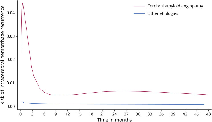
Objectives: Cerebral amyloid angiopathy (CAA)-associated lobar intracerebral hemorrhage (ICH) has a high risk of recurrence, but the underlying mechanisms remain uncertain. We, therefore, aimed to characterize patterns of recurrent ICH.

Methods: We investigated early recurrent ICH (≥1 recurrent ICH event within 90 days of the index event) and ICH clusters (≥2 ICH events within 90 days at any time point) in 2 large cohorts of consecutive patients with first-ever ICH and available MRI.

Results: In 682 included patients (median age 68 years, 40.3% female, median follow-up time 4.1 years), 18 (2.6%) had an early recurrent ICH, which was associated with higher age and CAA. In patients with probable CAA, the risk of early recurrent ICH was increased 5-fold within the first 3 months compared with during months 4-12 (hazard ratio 5.41, 95% CI 2.18-13.4) while no significant difference was observed in patients without CAA. In patients with an ICH cluster, we observed spatial clustering (recurrent ICH within close proximity of index ICH in 63.0%) and a tendency for multiple sequential hemorrhages (≥3 ICH foci within 3 months in 44.4%).

Discussion: Our data provide evidence of both temporal and spatial clustering of ICH in CAA, suggesting a transient and localized active bleeding-prone process.


Conflict of interest statement

The authors report no relevant disclosures. Go to Neurology.org/N for full disclosures.

Figures

Figure 1
Figure 1. Temporal Changes in Risk of Recurrent Intracerebral Hemorrhage
Figure 2
Figure 2. Neuroimaging Findings in 2 Exemplary Patients With ICH Clusters
(A) A patient in their seventies with index right parietal intracerebral hemorrhage. On index CT, 2 additional separate foci of convexity subarachnoid hemorrhage were visible (right parietal, left frontal). 48 days after the index ICH, the patient suffered a recurrent ICH in the right frontal lobe. On MRI after clinical worsening 6 days later, an additional large left frontal ICH was visible, as were disseminated cortical superficial siderosis and numerous lobar microbleeds. This patient did not have any further recurrent ICH over a follow-up period of 9 years (with several CT scans performed in the follow-up period). (B) Another patient in their seventies with right occipital intracerebral hemorrhage. T2* MRI sequences did not show additional chronic hemorrhagic lesions. 6 years later, the patient suffered a left frontal hemorrhage. After initial stabilization, MRI depicted bilateral parietal acute convexity subarachnoid hemorrhage and extensive cortical superficial siderosis. 53 days after the second ICH, the patient had a large right frontal ICH and died shortly thereafter. ICH = intracerebral hemorrhage.

References

    1. GBD 2019 Stroke Collaborators. Global, regional, and national burden of stroke and its risk factors, 1990-2019: a systematic analysis for the Global Burden of Disease Study 2019. Lancet Neurol. 2021;20(10):795-820. doi:10.1016/S1474-4422(21)00252-0 - DOI - PMC - PubMed
    1. Fogelholm R, Murros K, Rissanen A, Avikainen S. Long term survival after primary intracerebral haemorrhage: a retrospective population based study. J Neurol Neurosurg Psychiatry. 2005;76(11):1534-1538. doi:10.1136/jnnp.2004.055145 - DOI - PMC - PubMed
    1. Charidimou A, Imaizumi T, Moulin S, et al. . Brain hemorrhage recurrence, small vessel disease type, and cerebral microbleeds: a meta-analysis. Neurology. 2017;89(8):820-829. doi:10.1212/WNL.0000000000004259 - DOI - PMC - PubMed
    1. Fandler-Höfler S, Obergottsberger L, Ambler G, et al. . Association of the presence and pattern of MRI markers of cerebral small vessel disease with recurrent intracerebral hemorrhage. Neurology. 2023;101(8):e794-e804. doi:10.1212/WNL.0000000000207510 - DOI - PMC - PubMed
    1. Charidimou A, Peeters AP, Jäger R, et al. . Cortical superficial siderosis and intracerebral hemorrhage risk in cerebral amyloid angiopathy. Neurology. 2013;81(19):1666-1673. doi:10.1212/01.wnl.0000435298.80023.7a - DOI - PMC - PubMed

LinkOut - more resources