. 2024 Aug 16;15(1):7065.
doi: 10.1038/s41467-024-51290-0.

Motor innervation directs the correct development of the mouse sympathetic nervous system

Affiliations

Motor innervation directs the correct development of the mouse sympathetic nervous system

Alek G Erickson et al. Nat Commun. .

Abstract

The sympathetic nervous system controls bodily functions including vascular tone, cardiac rhythm, and the "fight-or-flight response". Sympathetic chain ganglia develop in parallel with preganglionic motor nerves extending from the neural tube, raising the question of whether axon targeting contributes to sympathetic chain formation. Using nerve-selective genetic ablations and lineage tracing in mouse, we reveal that motor nerve-associated Schwann cell precursors (SCPs) contribute sympathetic neurons and satellite glia after the initial seeding of sympathetic ganglia by neural crest. Motor nerve ablation causes mispositioning of SCP-derived sympathoblasts as well as sympathetic chain hypoplasia and fragmentation. Sympathetic neurons in motor-ablated embryos project precociously and abnormally towards dorsal root ganglia, eventually resulting in fusion of sympathetic and sensory ganglia. Cell interaction analysis identifies semaphorins as potential motor nerve-derived signaling molecules regulating sympathoblast positioning and outgrowth. Overall, central innervation functions both as infrastructure and regulatory niche to ensure the integrity of peripheral ganglia morphogenesis.


Conflict of interest statement

The authors declare no competing interests.

Figures

Fig. 1
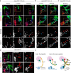
Fig. 1. SCP recruitment on motor nerves.
a Current model of sympathetic ganglia development from early stream of free neural crest migration. Free-NCCs (yellow) become primed toward autonomic fate (yellow-blue hybrid cells) as they approach the dorsal aorta region, where they differentiate into sympathetic neurons (blue) and coalesce to form the sympathetic ganglia chain. b Transverse sections of the same E10.5 Hb9-GFP embryo (representative of 3 embryos) from sacral to thoracic levels reveal the developmental progression of motor axons (Hb9-GFP labeling) exiting the ventral neural tube (outlined) and intersecting the stream of free NCCs (SOX10+). NCCs are recruited on motor nerves as SCPs (arrowheads) leaving a “gap” with freely migrating cells. Scatterplots of measurement of (c) the angle created by intersecting the line bisecting the NCC migratory stream, with the dorsoventral axis bisecting the neural tube (d) gap between nerve-associated SCP and the nearest free NCC, (e) mediolateral thickness of the NCC/SCP streams (distance between most medial and lateral SOX10+ cells just ventrolateral to the neural tube), perpendicular to (c). The red, blue, and green colors in (ce) represent measurements from individual E10.5 embryos (n = 3). Linear regression assessed correlation coefficients and p-values. f E10.5 transverse optical sections (representative of 3 embryos) show ITGA4+/SOX10+ SCP associated with axons (2H3, Neurofilament) at thoracic level, and free migrating ITGA4-/SOX10+ NCCs at sacral level. Single channels are magnified from the boxed regions in the merge image. The nerve bundle is outlined. g, h Transverse sections of Hb9-GFP embryos. At E10.5 (g), SCPs (SOX10+, gray) migrate along GFP+/TUJ1+ motor axons that form the ventral root before sensory axons (GFP-/TUJ1+) have begun to extend from the DRG (representative of 3 embryos). At E11.5 (h), SCPs are associated with both motor and sensory fascicles in the main nerve bundle (blue arrowheads). Along the white ramus connecting to sympathetic ganglia (PHOX2B+, blue), SCPs are largely associated with motor axons (red arrows), which form the bulk of the nerve (white arrowheads). The boxed regions are magnified in the right panels. Representative of 6 embryos. i Transverse sections representative of n = 5 E12.5 Hb9-GFP embryos with TRKA staining of sensory projections (red) co-extending with motor axons (GFP+, green). Individual channels are shown in the right panels. The boxed area, magnified in the bottom panel, shows the white ramus (arrowhead) formed predominantly by preganglionic motor axons, with minimal contribution from viscerosensory fibers. PHOX2B (blue) marks sympathetic ganglia. j Schematic showing equal distribution of SCPs (yellow) on motor (green) and sensory (red) fibers in axial and intercostal nerves, while SCPs migrating toward sympathetic chain ganglia (blue) are almost exclusively recruited on motor axons of the white ramus. DA dorsal aorta, DRG dorsal root ganglia, MN motor neurons, NCCs neural crest cells, NT neural tube, SCPs Schwann cell precursors, SG sympathetic chain ganglia, WR white ramus communicans. Scale bars: b: 100 μm; f: 50 μm; gi: 100 μm.
Fig. 2
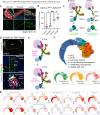
Fig. 2. SCPs are primed toward a sympathetic fate while migrating along motor nerves.
Transverse sections from E10.5 (a), E11.5 (b), and E12.5 (c) Hb9-GFP embryos immunostained for the NCC/SCP marker SOX10 (blue) and the sympathetic marker PHOX2B (red). Boxed regions are magnified in the corresponding bottom panels. Arrowheads point to committed PHOX2B+ SCPs associated with motor axons. Arrows point to PHOX2B- SCPs that may acquire glial fate within the ganglia. Representative of at least n = 4 embryos per stage. d Transverse view of E10.5 (upper panel) and E12.5 (bottom panel) Hb9-GFP embryo trunks immunostained for the early sympathetic marker PHOX2B (red) and differentiated sympathetic neuron marker TH (blue). The boxed regions are magnified to show individual PHOX2B (middle) and TH (right) staining. Arrowheads point to PHOX2B+/TH- SCPs associated with motor axons. The images are representative of at least 3 embryos per stage. e Schematic of motor nerves (green) assisting the late wave of SCP migration (yellow) towards sympathetic ganglia (blue). Motor axons represent a permissive substrate for autonomic priming (yellow-blue hybrid cells; PHOX2B+/TH-) but not for neuronal maturation (blue, TH+). SCPs that do not acquire PHOX2B expression might differentiate into satellite glial cells within sympathetic ganglia. DA dorsal aorta, DRG dorsal root ganglia, MN motor neurons, NCCs neural crest cells, NT neural tube, SCPs Schwann cell precursors, SG sympathetic chain ganglia, WR white ramus communicans. Scale bars: ac: 100 μm, 50 μm, 25 μm (from top to bottom panels); d: 100 µm.
Fig. 3
Fig. 3. Nerve-delivered SCPs contribute to sympathetic ganglia neurogenesis.
a Plp1-CreERT2; R26-YFP embryos traced by tamoxifen injection at E10.5 and harvested at E13.5. Immunostaining for TH and YFP on sagittal sections through sympathetic ganglia at different anatomical locations of the traced Plp1-CreERT2; R26-YFP embryos. b Quantifications of YFP+/TH+ cells traced at different levels of the sympathetic chain along the body axis. Colors represent individual embryos (n = 4). Mean ± SEM, one-way ANOVA and post hoc Tukey’s Multiple Comparison Test, (***) p = 0.001, (*) p = 0.0343, (ns) p = 0.6829. c Schematic of lineage tracing experiment. Induction of YFP expression in Plp1+ cells (yellow) at E10.5 results in differential contribution to sympathetic ganglia along different body segments. At brachial levels (left), in E10.5 embryos all YFP-labeled NCC derivatives are associated with nerves, revealing the extent of SCP contribution to sympathetic ganglia neurons (green cells in the ganglia at E13.5). At lumbar levels (right), YFP is induced in both nerve-associated SCPs and residual free-migrating NCCs at E10.5, resulting in mixed NCC/SCP contribution to sympathetic neurons (larger fraction of green cells in the ganglia at E13.5). d Transverse sections of Prss56-Cre; R26-Tomato E13.5 embryos immunostained for TH (red) and SOX10 (green) visualized alongside endogenous Tomato fluorescence (TOM, gray). Nuclei are in blue. (Top) TOM+ boundary caps adjacent to the neural tube (outlined). (Middle) BCC-derived SCPs (TOM+/SOX10+, arrowheads) in the ventral root (outlined). (Bottom) TH+/TOM+ traced cells (arrowheads) in sympathetic chain ganglia from the lower lumbar region (observed in n = 5/5 embryos). e Schematic showing the restricted expression of BCC markers, Prss56 and Egr2 in the boundary caps at E10.5 (upper panel) and tracing of BCC derivatives (purple) along nerves and sympathetic ganglia (bottom panel). f Combined UMAP embedding with color-coded scRNAseq clusters (top) and sample origin (bottom). Arrows show RNA velocity-determined transcriptional flows. g Feature plots showing expression of selected cell type marker genes. SG sympathetic ganglia, BCCs boundary cap cells, NCCs neural crest cells, NT neural tube, SCPs Schwann cell precursors. Scale bars: a: 50 μm; d: 100 μm (top), 50 μm (middle), 25 μm (bottom).
Fig. 4
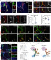
Fig. 4. Genetic ablation of motor neurons leads to ectopic autonomic priming along sensory nerves.
a Transverse sections showing complete loss of motor neuron cell bodies and axons (asterisks) in E11.5 Olig2-Cre; DTA embryos (right, n = 3) compared to control littermates (left, n = 2). Motor neurons are labeled with the cell-specific transgenic reporter MN(218-2)-GFP. In mutants, SCPs (SOX10+, red) migrate exclusively along sensory nerves (GFP-/TUJ1+). Individual labeling from the boxed regions are shown separately in the right panels. b Transverse sections at E12.5 showing mispatterning of viscerosensory projections (TRKA+/TUJ1+; arrowheads) in Olig2-Cre; DTA (bottom, n = 3) compared to controls (top, n = 3). PHOX2B (blue) identifies sympathetic ganglia. The boxed regions are magnified in middle and right panels. TRKA (gray) is shown separately in the right panels. c Transverse view of SCPs (SOX10, red) migrating on motor nerves (arrowheads) in E12.5 controls (left). SCP delivery to sympathetic ganglia (PHOX2B, TH) along the white ramus is interrupted (asterisks) in Olig2-Cre; DTA (right). SOX10 (gray) is shown separately in the right panels. d Area of glial cells (SOX10+ pixels) in sympathetic ganglia from E12.5 controls (black dots) and Olig2-Cre; DTA (blue dots). Mean (normalized to control) ± SEM, Unpaired two-sided t test (**) p = 0.0017; controls n = 4, mutants n = 4. e Sympathetic chain ganglia area (PHOX2B+ px) measured in transverse sections from E12.5 controls and Olig2-Cre; DTA. Mean (normalized to control) ± SEM, Unpaired two-sided t test (*) p = 0.0226; controls n = 7, mutants n = 7. f Time course of autonomic priming identified by PHOX2B+ cells associated with peripheral nerves (TUJ1+) outside sympathetic ganglia (arrowheads) in E10.5, E11.5, and E12.5 Olig2-Cre; DTA (bottom) and control (top) embryos. PHOX2B is shown separately in gray. Images are representative of at least 3 embryos per genotype, per stage. g Ectopic autonomic priming (PHOX2B+ cells) along sensory nerves (TRKA+/TUJ1+) away from the ganglia chain in E12.5 Olig2-Cre; DTA (bottom), but not in controls (top). The merged channels in the boxed regions are shown separately in the right panels. Average number of nerve-associated primed SCPs (PHOX2B+ cells outside the ganglia) (h) and distance from ganglia (i) in controls and Olig2-Cre; DTA. Mean ± SEM, Unpaired two-sided t test (*) p = 0.0136; (**) p = 0.0027; controls n = 3, mutants n = 3. j Schematics of autonomic priming (yellow-blue hybrid cells) on motor axons of the white ramus in the vicinity of the dorsal aorta in controls (left) compared to uncontrolled aberrant priming along sensory nerves far away from the sympathetic chain in motor nerve-ablated mutants (right). DA dorsal aorta, DRG dorsal root ganglia, MN motor neurons, NT neural tube, SCPs Schwann cell precursors, SG sympathetic chain ganglia, WR white ramus communicans. Scale bars: 100 µm.
Fig. 5
Fig. 5. Motor innervation controls sympathetic neuron differentiation, axonal projection and chain ganglia morphology.
Transverse sections at E11.5 (a) and E13.5 (b) showing misplaced sympathoblasts differentiating into sympathetic neurons (TH+, yellow) along sensory nerves (TUJ1+, cyan) and forming ectopic mini-ganglia in Olig2-Cre; DTA (arrowheads) but not in controls. SOX10 labels SCPs (magenta). TH staining is shown separately in the bottom panels (gray). Arrows point to fragmented sympathetic chain ganglia. Representative images of 6 embryos per genotype at E11.5 and 4 embryos per genotype at E13.5. c Dorsal view of a whole mount immunostaining for TH, Neurofilament (2H3), and vascular marker CD31 of E12.5 control embryo. The region encompassing the sympathetic chain visualized in (d) is outlined. d Dorsal view of a whole mount immunostaining for TH in control (left) and Olig2-Cre; DTA (right) E11.5 (top), E12.5 (middle), and E13.5 (bottom) embryos. Arrowheads point to sensory fiber-associated TH+ sympathetic neurons distant from the chain ganglia. Premature and aberrant extension of sympathetic fibers is visible in mutants. Representative images of at least n = 5 embryos per genotype at each stage. e Quantification of misplaced sensory nerve-attached sympathetic neurons per DRG between brachial and lumbar levels, averaged per embryo. Mean ± SEM, Unpaired two-sided t test (****) p < 0.0001; controls n = 3, mutants n = 3. f Quantification of misplaced TH+ cells outside the chain ganglia at E12.5, per embryo. Mean ± SEM, Unpaired two-sided t test (**) p = 0.0066; controls n = 3, mutants n = 4. g Distribution of the distances between misplaced TH+ cells and the borders of the chain ganglia. Each color of dot represents a different embryo (n = 3 controls, n = 4 mutants). Sagittal view of thoracic (h) and lumbar (i) regions from whole mount TH immunostaining of E12.5 (top) and E13.5 (bottom) Olig2-Cre; DTA embryos (right) and control littermates (left). Yellow arrowheads point to fragmented sympathetic chain in mutants; red arrowheads indicate abnormal sympathetic axon growth. Representative images of at least n = 5 embryos per genotype at each stage. j Volume measurements of sympathetic chain ganglia, cervical ganglia, and adrenal/paraganglia. Datapoints represent the average volume of ganglia from individual embryos (n = 3 control, n = 4 mutant). Mean ± SEM, one-way ANOVA using Šidák correction for multiple comparisons (**) p = 0.0032; (*) p = 0.0251; ns: p = 0.9815. k Oblique view of whole mount immunostaining for TH, Neurofilament (2H3), and vascular marker CD31 of E12.5 control embryo. The outlined forelimb region is shown in (l). l Forelimbs of E11.5 (top) and E12.5 (bottom) controls (left) and Olig2-Cre; DTA (right) littermates. Red arrowheads point to ectopic TH+ neurons extending axons aberrantly into the limb. m Length of TH+ sympathetic axons innervating the forelimb in control littermates vs Olig2-Cre; DTA embryos (at E11, controls n = 4, mutants n = 3; at E12.5, controls n = 3, mutants n = 4). Mean ± SEM, one-way ANOVA using Šidák correction for multiple comparisons (***) p = 0.0003; (*) p = 0.0118. n Schematic showing both ectopic and normally positioned sympathetic neurons (blue) projecting prematurely and along inappropriate paths in association with sensory fibers in motor-ablated mutants. The sympathetic chain is fragmented. DRG dorsal root ganglia, MN motor neurons, NT neural tube, OZ organ of Zuckerkandl, SCPs Schwann cell precursors, SG sympathetic chain ganglia. Scale bars: a, b: 100 µm, c: 500 µm; d: 200 µm (top and middle), 300 µm (bottom); h, i: 100 µm (top), 200 µm (bottom); k: 500 µm; l: 200 µm (top), 300 µm (bottom).
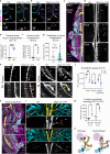
Fig. 6
Fig. 6. Motor nerves serve as insulators to prevent abnormal intermixing of sympathetic and sensory ganglia.
a Transverse embryo sections at E15.5 showing fusion of sympathetic (TH+, magenta) and dorsal root ganglia (TRKA+, cyan) in Olig2-Cre; DTA (right), but not in control littermates (left). TRKA and TH are shown separately in middle and bottom panels, respectively. b Distance between sympathetic and dorsal root ganglia in Olig2-Cre; DTA and controls. Mean ± SEM, Unpaired two-sided t test (****) p < 0.0001; controls n = 9, mutants n = 7. c Immunostaining for TH (magenta), SOX10 (yellow), and 2H3 (cyan) in sagittal sections of Olig2-Cre; DTA embryos and control littermates. The dorsal root ganglion is outlined with a dotted line. Boxed regions are magnified in the insets showing interspersed sensory neurons in sympathetic ganglia (white arrowheads). Yellow arrowheads point to fusion of DRG and sympathetic ganglia in mutants. d Fraction of ganglia pairs showing aberrant organization such as misplaced sympathoblasts around the sensory ganglia, or misplaced sensory neurons in the sympathetic ganglia. Mean ± SEM, Unpaired two-sided t test (**) p = 0.0046; controls n = 3, mutants n = 3. e Average cross-sectional area of DRGs per embryo. Mean ± SEM, Unpaired two-sided t test (*) p = 0.0196; controls n = 4, mutants n = 5. f Transverse view of fragmented DRG (TRKA+, cyan) (arrowheads) in E18.5 Olig2-Cre; DTA (bottom) but not controls (top). TH (magenta) and TUJ1 (yellow) mark sympathetic projections. Representative images of n = 4 controls and n = 5 mutants. g Schematic showing intermixing of sympathetic (blue) and sensory (red) ganglia in the absence of motor nerves (green). NT neural tube, SG sympathetic ganglia, DRG dorsal root ganglia, MN motor neurons. Scale bars: a: 100 µm; c: 200 µm (insets 50 µm); f: 100 µm.

References

    1. Wehrwein, E. A., Orer, H. S. & Barman, S. M. Overview of the anatomy, physiology, and pharmacology of the autonomic nervous system. Compr. Physiol. 6, 1239–1278 (2016). - PubMed
    1. Ernsberger, U. & Rohrer, H. Sympathetic tales: subdivisons of the autonomic nervous system and the impact of developmental studies. Neural Dev.13, 20 (2018). 10.1186/s13064-018-0117-6 - DOI - PMC - PubMed
    1. McCorry, L. K. Physiology of the autonomic nervous system. Am. J. Pharm. Educ.71, 78–78 (2007). 10.5688/aj710478 - DOI - PMC - PubMed
    1. Newbern, J. M. Molecular control of the neural crest and peripheral nervous system development. Curr. Top. Dev. Biol.111, 201–231 (2015). 10.1016/bs.ctdb.2014.11.007 - DOI - PMC - PubMed
    1. Serbedzija, G. N., Bronner-Fraser, M. & Fraser, S. E. A vital dye analysis of the timing and pathways of avian trunk neural crest cell migration. Development106, 809–816 (1989). 10.1242/dev.106.4.809 - DOI - PubMed