Multicenter Study
. 2024 Nov 14;83(12):1762-1772.
doi: 10.1136/ard-2024-225868.

Plasma proteome profiling in giant cell arteritis

Affiliations
Multicenter Study

Plasma proteome profiling in giant cell arteritis

Kevin Y Cunningham et al. Ann Rheum Dis. .

Abstract

Objectives: This study aimed to identify plasma proteomic signatures that differentiate active and inactive giant cell arteritis (GCA) from non-disease controls. By comprehensively profiling the plasma proteome of both patients with GCA and controls, we aimed to identify plasma proteins that (1) distinguish patients from controls and (2) associate with disease activity in GCA.

Methods: Plasma samples were obtained from 30 patients with GCA in a multi-institutional, prospective longitudinal study: one captured during active disease and another while in clinical remission. Samples from 30 age-matched/sex-matched/race-matched non-disease controls were also collected. A high-throughput, aptamer-based proteomics assay, which examines over 7000 protein features, was used to generate plasma proteome profiles from study participants.

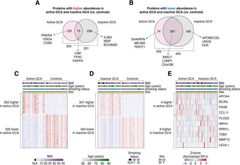
Results: After adjusting for potential confounders, we identified 537 proteins differentially abundant between active GCA and controls, and 781 between inactive GCA and controls. These proteins suggest distinct immune responses, metabolic pathways and potentially novel physiological processes involved in each disease state. Additionally, we found 16 proteins associated with disease activity in patients with active GCA. Random forest models trained on the plasma proteome profiles accurately differentiated active and inactive GCA groups from controls (95.0% and 98.3% in 10-fold cross-validation, respectively). However, plasma proteins alone provided limited ability to distinguish between active and inactive disease states within the same patients.

Conclusions: This comprehensive analysis of the plasma proteome in GCA suggests that blood protein signatures integrated with machine learning hold promise for discovering multiplex biomarkers for GCA.

Keywords: Giant Cell Arteritis; Machine Learning; Vasculitis.


Conflict of interest statement

Competing interests: None declared.

Figures

Figure 1
Figure 1. Analysis of plasma proteome profiles in patients with giant cell arteritis (GCA) and controls. (A) Study design overview: Plasma samples were collected from patients with GCA in two instances: during an active disease state (active GCA group, n=30) and while in clinical remission (inactive GCA group, n=30). Concurrently, plasma samples from controls (n=30) were also collected. Proteome profiling of 7289 proteins in all 90 samples was performed using the SomaScan Assay version 4 (SomaLogic), and these profiles were subsequently used in downstream statistical analyses. (B) Principal component analysis (PCA) on the plasma proteome profiles, where each point on the scatter plot represents an individual sample. Box-and-whisker plots displayed on the top and to the right of the plot represent the distribution of principal component 1 (PC1) and principal component 2 (PC2) values, respectively. (C) Clinical and demographic factors associated with PC1 values. Bar height represents the –log10 transformation of the p value of the association, with the significance threshold indicated in blue (p=0.05). Additionally, the size of the data points on the plot corresponds to the R2 value from each linear regression model. BMI, body mass index; PGA, physician global assessment; CRP, C reactive protein.
Figure 2
Figure 2. Differentially abundant plasma proteins between study groups. (A, B) Of the 7289 measured plasma proteins, 984 showed differential abundance between GCA groups (ie, active or inactive GCA) and controls. In active GCA, the abundance of 202 and 335 proteins were higher and lower compared with controls, respectively. Conversely, in inactive GCA, the abundance of 331 and 450 proteins were higher and lower than in controls, respectively. Heatmaps showing the Z-score-normalised relative fluorescence unit (RFU) abundances of differentially abundant proteins between (C) active GCA and controls (537 proteins); (D) inactive GCA and controls (781 proteins) and (E) active and inactive GCA (10 proteins). In each heatmap, the columns represent study participants while the rows correspond to proteins. Demographic variables (BMI, age, smoking status, and sex) of each study subject are shown at the top of each heatmap. BMI, body mass index; GCA, giant cell arteritis.
Figure 3
Figure 3. Gene ontology (GO) enrichment analysis of differentially abundant plasma proteins in giant cell arteritis. (A, B) Top 20 enriched GO biological processes of proteins with significantly higher abundance in active (or inactive) GCA compared with controls. Proteins with higher abundance in either GCA group are mostly linked to immune processes. (C, D) Top 20 enriched biological processes of proteins with significantly lower abundance in active (or inactive) GCA than in controls. Most of these proteins were enriched in functions related to metabolism, cell signalling and transport. GO biological processes (located to the left of each bar graph) are arranged in descending order based on significance, represented by the –log10 of the p values obtained from a modified one-tailed Fisher’s exact test. Longer bars indicate greater significance while shading illustrates fold-enrichment. GCA, giant cell arteritis.
Figure 4
Figure 4. Plasma protein correlations with physician global assessment (PGA) and C reactive protein (CRP) in patients with active giant cell arteritis. (A) Five proteins displayed positive correlations with PGA scores (Spearman’s ρ>0.4 and p<0.05 in MLRMs), whereas 11 proteins exhibited negative correlations (Spearman’s ρ<–0.4 and p<0.05 in MLRMs). (B) 10 proteins were positively correlated with blood CRP levels (measured in mg/dL) while 11 proteins showed negative correlations. MLRMs, multiple linear regression models.
Figure 5
Figure 5. Classification accuracy of random forest classifiers with varying sizes of input feature sets. Pink bars indicate the accuracy in classifying active giant cell arteritis (GCA) versus controls, and grey represent accuracy in classifying inactive GCA versus controls. This process involved preselecting a specified number of input plasma protein features. Different numbers of top-selected features were chosen in each training set fold, ranging from 10 to 250. These selected features were then applied in a random forest classifier, with accuracy assessed on the test set across all ten folds of cross-validation.

References

    1. Pugh D, Karabayas M, Basu N, et al. Large-vessel vasculitis. Nat Rev Dis Primers. 2022;7:1–23. doi: 10.1038/s41572-021-00327-5. - DOI - PMC - PubMed
    1. Kermani TA, Schmidt J, Crowson CS, et al. Utility of erythrocyte sedimentation rate and C-reactive protein for the diagnosis of giant cell arteritis. Semin Arthritis Rheum. 2012;41:866–71. doi: 10.1016/j.semarthrit.2011.10.005. - DOI - PMC - PubMed
    1. van der Geest KSM, Abdulahad WH, Rutgers A, et al. Serum markers associated with disease activity in giant cell arteritis and polymyalgia rheumatica. Rheumatol (Oxford) 2015;54:1397–402. doi: 10.1093/rheumatology/keu526. - DOI - PubMed
    1. Burja B, Feichtinger J, Lakota K, et al. Utility of serological biomarkers for giant cell arteritis in a large cohort of treatment-naïve patients. Clin Rheumatol. 2019;38:317–29. doi: 10.1007/s10067-018-4240-x. - DOI - PubMed
    1. Wadström K, Jacobsson LTH, Mohammad AJ, et al. Analyses of plasma inflammatory proteins reveal biomarkers predictive of subsequent development of giant cell arteritis: a prospective study. Rheumatology (Oxford) 2023;62:2304–11. doi: 10.1093/rheumatology/keac581. - DOI - PMC - PubMed

Publication types