. 2024 Aug 17;17(1):167.
doi: 10.1186/s13048-024-01484-3.

Granulosa cell insight: unraveling the potential of menstrual blood-derived stem cells and their exosomes on mitochondrial mechanisms in polycystic ovary syndrome (PCOS)

Affiliations

Granulosa cell insight: unraveling the potential of menstrual blood-derived stem cells and their exosomes on mitochondrial mechanisms in polycystic ovary syndrome (PCOS)

Mahna Mansoori et al. J Ovarian Res. .

Abstract

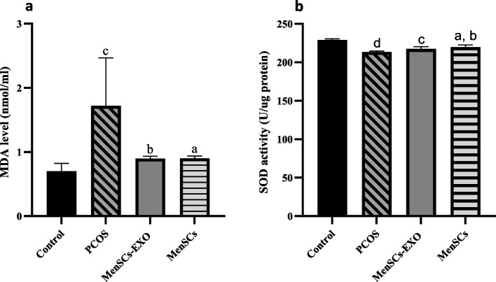
Background: Polycystic ovary syndrome (PCOS) presents a significant challenge in women's reproductive health, characterized by disrupted folliculogenesis and ovulatory dysfunction. Central to PCOS pathogenesis are granulosa cells, whose dysfunction contributes to aberrant steroid hormone production and oxidative stress. Mitochondrial dysfunction emerges as a key player, influencing cellular energetics, oxidative stress, and steroidogenesis. This study investigates the therapeutic potential of menstrual blood-derived stem cells (MenSCs) and their exosomes in mitigating mitochondrial dysfunction and oxidative stress in PCOS granulosa cells.

Methods: Using a rat model of PCOS induced by letrozole, granulosa cells were harvested and cultured. MenSCs and their exosomes were employed to assess their effects on mitochondrial biogenesis, oxidative stress, and estrogen production in PCOS granulosa cells.

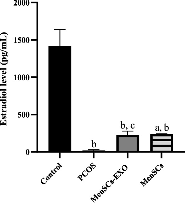
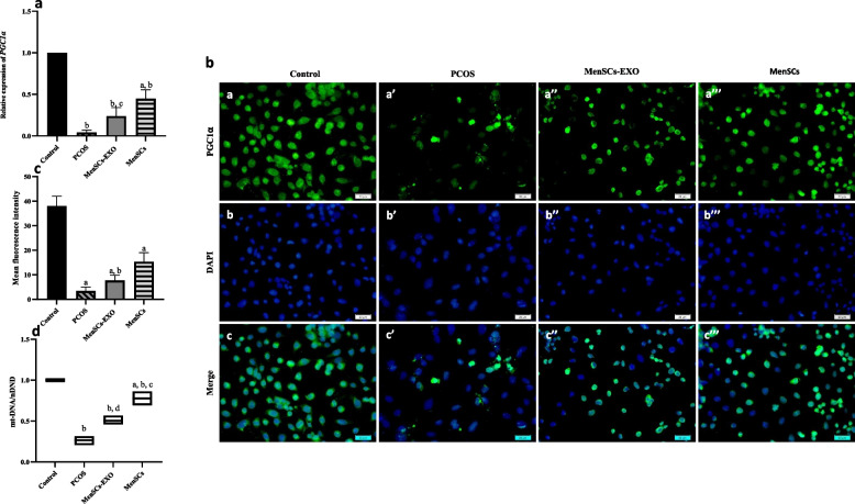
Results: Results showed diminished mitochondrial biogenesis and increased oxidative stress in PCOS granulosa cells, alongside reduced estrogen production. Treatment with MenSCs and their exosomes demonstrated significant improvements in mitochondrial biogenesis, oxidative stress levels, and estrogen production in PCOS granulosa cells. Further analysis showed MenSCs' superior efficacy over exosomes, attributed to their sustained secretion of bioactive factors. Mechanistically, MenSCs and exosomes activated pathways related to mitochondrial biogenesis and antioxidative defense, highlighting their therapeutic potential for PCOS.

Conclusions: This study offers insights into granulosa cells mitochondria's role in PCOS pathogenesis and proposes MenSCs and exosomes as a potential strategy for mitigating mitochondrial dysfunction and oxidative stress in PCOS. Further research is needed to understand underlying mechanisms and validate clinical efficacy, presenting promising avenues for addressing PCOS complexity.

Keywords: Exosomes; Granulosa cells; Menstrual blood-derived stem cells (MenSCs); Mitochondrial biogenesis; Polycystic ovary syndrome (PCOS).


Conflict of interest statement

The authors declare no competing interests.

Figures

Fig. 1
Fig. 1
Photomicrographs illustrate the characteristic cytology of vaginal smears obtained from control rats. In rats, the cycle stages were classified as proestrus (PRO), characterized by a predominant population of nucleated epithelial cells (green arrow); estrus (EST), distinguished by cornified cells (white arrow); metestrus (MET), identified by the presence of both cornified epithelial cells (white arrow), nucleated epithelial cells (green arrow) and leukocytes (black arrow); and the diestrus stage (DIST), where leukocytes predominate alongside nucleated epithelial cells (green arrow).” 400 × magnification; bars = 50 μm
Fig. 2
Fig. 2
Histological photomicrographs of rat ovaries stained with Mason trichrome. a PCOS group; Displays the presence of follicular cysts (FC) and the absence of normal follicle development. b Control group; Shows normal ovarian structures, including Corpus luteum (CL), Primary Follicle (PF), Secondary Follicle (SF), and Antral Follicle (AF).; 40 × magnification; bars = 200 μm
Fig. 3
Fig. 3
Morphological characteristic and Follicle-stimulating hormone receptor (FSHR) expression assay of granulosa cells In-vitro. a The morphological characteristics of granulosa cells were visualized using an inverted microscope. The cells exhibited an epithelial-like morphology, with a flat and polygonal shape. Nuclei appeared round. 100 × magnification; bars = 100 μm. b Immunofluorescence staining of FSHR (green) was performed in granulosa cells obtain from rat ovaries. The nuclei were stain with DAPI (blue). 400 × magnification; bars = 20 μm
Fig. 4
Fig. 4
Morphological characteristic and identification of Menstrual Blood-Derived Mesenchymal Stem Cells (MenSCs). a Morphology of passage 4–6 MenSCs observed under an inverted microscope showing spindle-shaped cells. 40 × magnification; bars = 200 μm. b Surface marker expression profile of MenSCs analyzed by flow cytometry; these cells were positive for CD105, CD90, and negative for CD34 and CD45, confirming their mesenchymal stem cell identity
Fig. 5
Fig. 5
Characterization of exosomes derived from Menstrual Blood-Derived Mesenchymal Stem Cells (MenSCs-EXO). a Morphology of MenSCs-EXO observed under a transmission electron microscope demonstrating a heterogenous size mixture of cup-shaped vesicles ranging from 50–150 nm. b Particle diameter distribution range of MenSCs-EXO measured by Dynamic Light Scattering (DLS) in triplicate revealed an average diameter of 59 nm. c Western blot analysis demonstrated the presence of exosome markers CD63 and CD9 in MenSCs-EXO
Fig. 6
Fig. 6
Uptake of PKH67-labeled MenSCs-EXO by granulosa cells In-vitro. Exosomes, purified from condition media of MenSCs culture, were labeled with PKH67 dye and co-cultured with primary culture of granulosa cells for 24 h. Green fluorescence indicates the successful uptake of PKH67-labeled MenSCs-EXO by the granulosa cells, while blue fluorescence represents nuclear staining with Hoechst. 100 × magnification; bars = 100 μm
Fig. 7
Fig. 7
Graph of MTT Viability Assay Conducted on PCOS Granulosa Cells Exposed to Different Concentrations of MenSCs-Exo Over a 24-h Period. The concentrations of 8 and 16 μg/mL MenSCs-exo significantly increased proliferation compared to non-treated cells. Data are presented as mean ± standard deviation. a = P ≤ 0.0006 vs non-treated cells and b = P ≤ 0.0001 vs non-treated cells
Fig. 8
Fig. 8
Oxidative Stress Parameters in Granulosa Cells following treatment with MenSCs and MenSCs- EXO. a MDA concentrations in culture medium of different groups showing higher levels in PCOS compared to control, with reductions following treatment with MenSCs and MenSCs-EXO. Data are presented as mean ± standard deviation. a = P ≤ 0.016 vs PCOS; b = P ≤ 0.015 vs PCOS; c = P ≤ 0.003 vs Control. b SOD Activity in cell lysates, showing a significant increase in MenSCs-treated cells compared to PCOS, while MenSCs-EXO treatment did not significantly change SOD activity. Data are presented as mean ± standard deviation. a = P ≤ 0.011 vs PCOS; b = P ≤ 0.001 vs Control; c = P ≤ 0.002 vs Control and d = P ≤ 0.0001 vs Control
Fig. 9
Fig. 9
Estradiol level in culture medium of different groups following treatment with MenSCs-EXO and MenSCs. Estradiol levels were significantly lower in PCOS compared to control. Treatment with both MenSCs and MenSCs-EXO led to a substantial increase in estradiol production in PCOS granulosa cells. Data are presented as mean ± standard deviation. a = P ≤ 0.007 vs PCOS; b = P ≤ 0.0001 vs Control + PCOS and c = P ≤ 0.012 vs PCOS
Fig. 10
Fig. 10
Mitochondrial Biogenesis Status. a Relative Expression Level of Gene PGC1α by real time PCR. Results were normalized at first with GAPDH and then to the Control. Data are presented as mean ± standard deviation. a = P ≤ 0.002 vs MenSCs-EXO + PCOS + Control; b = P ≤ 0.0001 vs MenSCs-EXO + PCOS + Control and c = P ≤ 0.004 vs PCOS. b Immunofluorescence Staining of PGC1α (green) was performed in granulosa cells. The nuclei were stain with DAPI (blue). 400 × magnification; bars = 20 μm. c Semi-quantification of Mean Fluorescence Intensity of PGC1α in granulosa Cells following treatment with MenSCs-EXO and MenSCs. Data are presented as mean ± standard deviation. a = P ≤ 0.0001 vs MenSCs-EXO + PCOS + Control; b = P ≤ 0.044 vs PCOS. d Relative mtDNA Copy Number in Granulosa Cells of different groups were measured by quantitative real-time PCR (qPCR) and reported as a ratio of mitochondrial DNA (mt-DNA) to the nuclear DNA (nDNA). Data are presented as mean. a = P ≤ 0.001 vs MenSCs-EXO; b = P ≤ 0.0001 vs PCOS + Control and c = P ≤ 0.006 vs Control and d = P ≤ 0.004 vs PCOS

References

    1. Lai Q, et al. Oxidative stress in granulosa cells contributes to poor oocyte quality and IVF-ET outcomes in women with polycystic ovary syndrome. Front Med. 2018;12:518–24. 10.1007/s11684-017-0575-y - DOI - PubMed
    1. Rooda I, et al. Cellular, extracellular and extracellular vesicular miRNA profiles of pre-ovulatory follicles indicate signaling disturbances in polycystic ovaries. Int J Mol Sci. 2020;21(24):9550. 10.3390/ijms21249550 - DOI - PMC - PubMed
    1. Deng Y, et al. Whole genome transcriptomic analysis of ovary granulosa cells revealed an anti-apoptosis regulatory gene DLGAP5 in polycystic ovary syndrome. Front Endocrinol. 2022;13:781149.10.3389/fendo.2022.781149 - DOI - PMC - PubMed
    1. Chauvin S, Cohen-Tannoudji J, Guigon CJ. Estradiol signaling at the heart of folliculogenesis: its potential deregulation in human ovarian pathologies. Int J Mol Sci. 2022;23(1):512. 10.3390/ijms23010512 - DOI - PMC - PubMed
    1. Tu J, et al. The role of microRNAs in ovarian granulosa cells in health and disease. Front Endocrinol. 2019;10:174.10.3389/fendo.2019.00174 - DOI - PMC - PubMed

LinkOut - more resources