. 2024 Aug 17;15(1):7083.
doi: 10.1038/s41467-024-50692-4.

α-Synuclein oligomers form by secondary nucleation

Affiliations

α-Synuclein oligomers form by secondary nucleation

Catherine K Xu et al. Nat Commun. .

Abstract

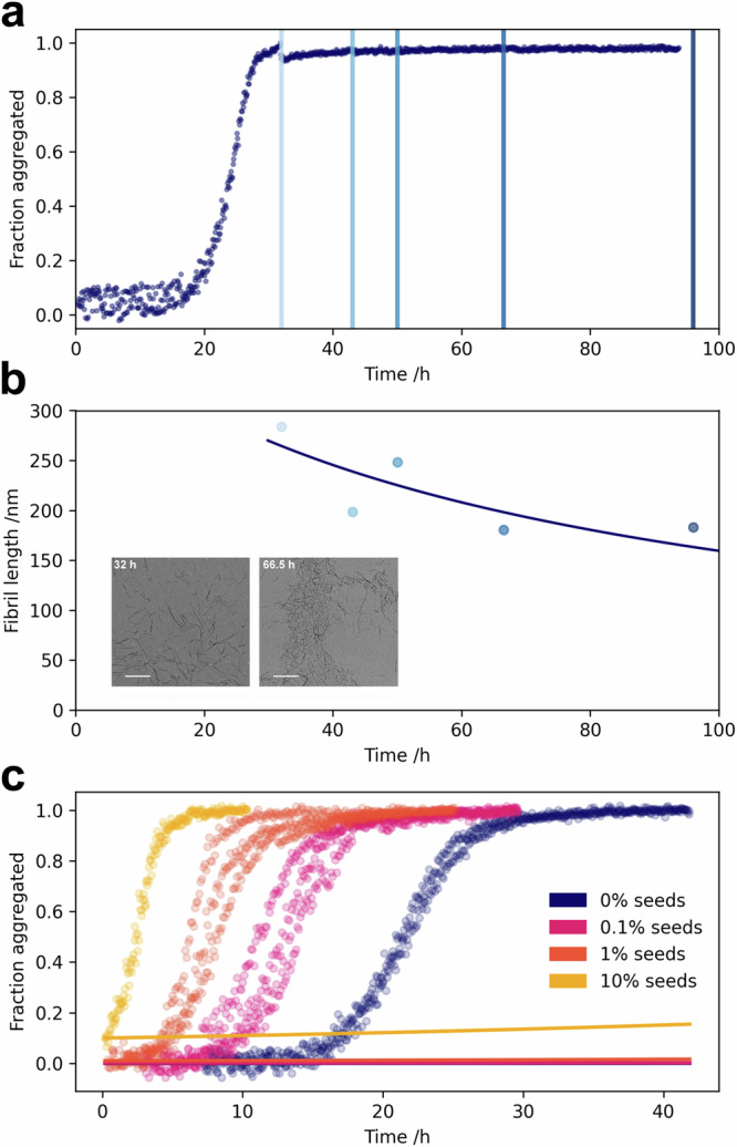
Oligomeric species arising during the aggregation of α-synuclein are implicated as a major source of toxicity in Parkinson's disease, and thus a major potential drug target. However, both their mechanism of formation and role in aggregation are largely unresolved. Here we show that, at physiological pH and in the absence of lipid membranes, α-synuclein aggregates form by secondary nucleation, rather than simple primary nucleation, and that this process is enhanced by agitation. Moreover, using a combination of single molecule and bulk level techniques, we identify secondary nucleation on the surfaces of existing fibrils, rather than formation directly from monomers, as the dominant source of oligomers. Our results highlight secondary nucleation as not only the key source of oligomers, but also the main mechanism of aggregate formation, and show that these processes take place under conditions which recapitulate the neutral pH and ionic strength of the cytosol.


Conflict of interest statement

At the time of initial submission, Georg Meisl and Alexander J Dear were employees of Wavebreak Therapeutics (formerly Wren Therapeutics). Michele Vendruscolo, Sara Linse, and Tuomas PJ Knowles are co-founders of Wavebreak Therapeutics (formerly Wren Therapeutics). The remaining Authors declare no competing interests.

Figures

Fig. 1
Fig. 1. Using microfluidic free-flow electrophoresis with single molecule detection, we were able to fractionate α-synuclein aggregation mixtures and determine oligomer dynamics.
By using chemical kinetics we determined that secondary nucleation on fibril surfaces is the dominant mechanism of both α-synuclein oligomers and new fibrils.
Fig. 2
Fig. 2. Secondary processes are involved in the aggregation of α-synuclein.
The aggregation kinetics of AlexaFluor-488-labelled α-synuclein were followed by aggregation-induced quenching of the AlexaFluor-488 dye (Fig. S7). The aggregation kinetics of the labeled and unlabelled α-synuclein are the same (Fig. S6 and Table S1). The dependence of α-synuclein aggregation kinetics on the absence and presence of varying concentrations of fibrillar seeds. Data were fitted to kinetic models in the absence (a) and presence (b) of secondary processes. The data are only consistent with a model that includes the fibril-catalyzed formation of new fibrils.
Fig. 3
Fig. 3. Fragmentation does not account for α-synuclein fibril proliferation.
Fibrils were withdrawn from an aggregation reaction at the indicated timepoints in the plateau phase of the aggregation mixture (a) and imaged by TEM (b), inset, scale bar = 1 μM). b The mean lengths of fibrils were determined from at least 8 TEM images containing a minimum of 650 fibrils in total, and fitted to kinetic models to determine the fragmentation rate, finding a very low value of 0.01 h−1. c The kinetic data were then fitted with fragmentation as the mechanism of fibril amplification, with the fragmentation rate constant fixed to the value determined in (b).
Fig. 4
Fig. 4. α-Synuclein oligomers form by secondary nucleation.
Aggregation mixtures at various time points throughout the reaction were centrifuged (21, 130 × g, 10 min, 20 °C) to remove large fibrillar aggregates and the oligomer content of the resulting supernatant studied by μFFE at the single molecule level (a). Kinetics of fibrillar α-synuclein in unseeded (blue) and seeded (red, 1% seeds) aggregation reactions, measured by fluorescence quenching, are shown alongside the fitted model (b). The relative oligomer mass concentrations were determined (c) and fitted to a model in which oligomers can form via both primary nucleation from monomers and secondary nucleation on fibril surfaces (model details in SI). X-axis error bars represent the time range over which data were averaged, which corresponds to the standard deviation of the aggregation half times. Where present, y-axis error bars represent the standard error of the mean oligomer mass concentrations from up to 8 biological replicates; points without y-axis error bars represent a single sample.
Fig. 5
Fig. 5. Secondary nucleation proceeds under quiescent, physiological conditions.
Secondary nucleation involves the formation of oligomers on fibril surfaces, followed by their release into solution (a). α-Synuclein was aggregated under both quiescent and shaking conditions (b, c), and the oligomer populations investigated. The relative oligomer mass concentrations of quiescent seeded (1%) aggregation reactions at the half-time (indicated by the dotted line in (c) before and after shaking for 10 minutes at 200 rpm are shown alongside the oligomer concentration during the plateau phase of the reaction under shaking (d). Example timetraces of photon count rates are shown for quiescent seeded (1%) aggregation reactions before (e) and after (f) shaking.
Fig. 6
Fig. 6. CSF from Parkinson’s disease patients catalyses oligomer formation.
a Aggregation kinetics of α-synuclein aggregation in the presence of 4% v/v pooled CSF from Parkinson’s disease patients and a healthy cohort. The extracted lag times (time taken to reach 25% aggregation) are shown alongside unseeded and seeded (1% mass concentration) reactions in the absence of CSF (inset). b, c Example photon count timetraces from aggregation reactions seeded by PD CSF (b) and in vitro-generated seeds (c). Oligomers from the PD CSF-seeded reaction were investigated by μFFE at around 30% aggregation (b) and found to have similar biophysical properties to a corresponding sample from the in vitro seeded reaction (c).

References

    1. Chiti, F. & Dobson, C. M. Protein misfolding, amyloid formation, and human disease: a summary of progress over the last decade. Annu. Rev. Biochem.86, 27–68 (2017). 10.1146/annurev-biochem-061516-045115 - DOI - PubMed
    1. Soto, C. Unfolding the role of protein misfolding in neurodegenerative diseases. Nat. Rev. Neurosci.4, 49–60 (2003). 10.1038/nrn1007 - DOI - PubMed
    1. Spillantini, M. G. et al. Alpha-synuclein in lewy bodies. Nature388, 839–840 (1997). 10.1038/42166 - DOI - PubMed
    1. Flagmeier, P. et al. Mutations associated with familial parkinson’s disease alter the initiation and amplification steps of α-synuclein aggregation. Proc. Natl Acad. Sci.113, 10328–10333 (2016). 10.1073/pnas.1604645113 - DOI - PMC - PubMed
    1. Bell, R. et al. Effects of n-terminal acetylation on the aggregation of disease-related α-synuclein variants. J. Mol. Biol.435, 167825 (2023). 10.1016/j.jmb.2022.167825 - DOI - PubMed

LinkOut - more resources