

Presentation of mitral valve cleft with concurrent atrial septal defect and ventricular septal defect detected by three-dimensional transesophageal echocardiography: a case report
- PMID: 39153999
- PMCID: PMC11330598
- DOI: 10.1186/s13256-024-04704-y
Presentation of mitral valve cleft with concurrent atrial septal defect and ventricular septal defect detected by three-dimensional transesophageal echocardiography: a case report
Abstract
Background: Cleft in the mitral valve leaflet is a primary cause of congenital mitral regurgitation, stemming from developmental anomalies in the mitral valve and frequently associated with other congenital heart defects. Concurrent presence of cleft in mitral valve leaflet with atrial septal defect and ventricular septal defect is relatively rare. Echocardiography, especially transesophageal echocardiography, is essential in diagnosing cleft mitral valve leaflet and related congenital heart defects, providing critical, detailed imagery for accurate assessment. This study presents a young female patient whose anterior mitral cleft, along with atrial septal defect and ventricular septal defect, was revealed through three-dimensional transesophageal echocardiography.
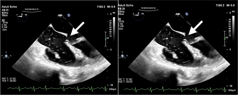
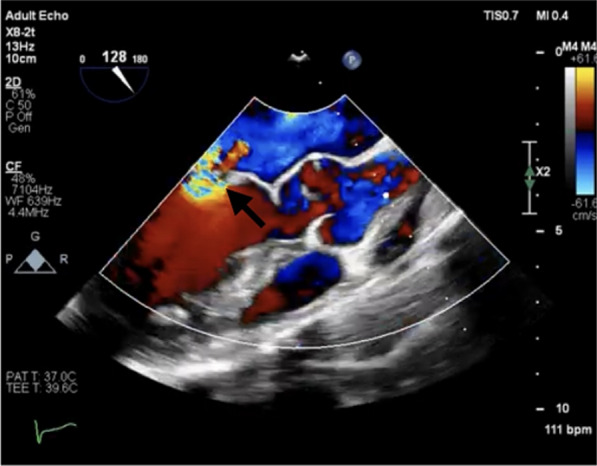
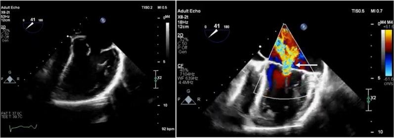
Case presentation: A 25-year-old Iranian female, experiencing progressive dyspnea and diminished physical capacity over 3 months, was referred to our hospital. Initial examination and transthoracic echocardiography indicated severe mitral regurgitation. Further evaluation with transesophageal echocardiography corroborated these findings and identified a cleft in the anterior mitral valve leaflet, coupled with mild left ventricular enlargement and significant left atrial enlargement. The complexity of the patient's condition was heightened by the diagnosis of cleft mitral valve leaflet in conjunction with atrial septal defect and ventricular septal defect, showing the complex nature of congenital defects.
Conclusion: This case emphasizes the critical role of transthoracic echocardiography in diagnosing cleft of mitral valve leaflet and associated cardiac anomalies, showcasing its superiority over transthoracic echocardiography for detailed visualization of cardiac structures. The identification of multiple congenital defects highlights the necessity for a comprehensive diagnostic approach to manage and treat patients with complex congenital heart diseases effectively. Future research should aim to refine diagnostic methodologies to enhance patient outcomes for cleft of mitral valve leaflets and related congenital conditions.
Keywords: Atrial septal defect; Case report; Echocardiography; Mitral valve cleft; Ventricular septal defect.
© 2024. The Author(s).
Conflict of interest statement
The authors declare that they have no competing interests.
Figures

References
-
- Pavlicek J, Gruszka T, Kapralova S, Prochazka M, Silhanova E, Kaniova R, et al. Associations between congenital heart defects and genetic and morphological anomalies. The importance of prenatal screening. Biomed Pap Med Fac Univ Palacky Olomouc Czech Repub. 2019;163(1):67–74. 10.5507/bp.2018.049 - DOI - PubMed
Publication types
MeSH terms
LinkOut - more resources
Full Text Sources
