. 2024 Aug 17;15(1):7092.
doi: 10.1038/s41467-024-51259-z.

Structure of the human TIP60 complex

Affiliations

Structure of the human TIP60 complex

Ke Chen et al. Nat Commun. .

Abstract

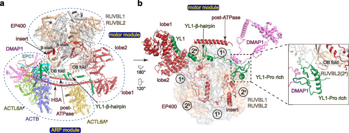
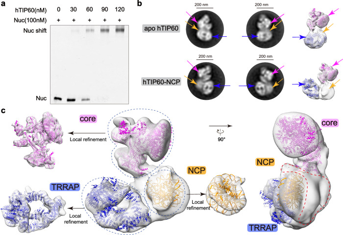
Mammalian TIP60 is a multi-functional enzyme with histone acetylation and histone dimer exchange activities. It plays roles in diverse cellular processes including transcription, DNA repair, cell cycle control, and embryonic development. Here we report the cryo-electron microscopy structures of the human TIP60 complex with the core subcomplex and TRRAP module refined to 3.2-Å resolution. The structures show that EP400 acts as a backbone integrating the motor module, the ARP module, and the TRRAP module. The RUVBL1-RUVBL2 hexamer serves as a rigid core for the assembly of EP400 ATPase and YL1 in the motor module. In the ARP module, an ACTL6A-ACTB heterodimer and an extra ACTL6A make hydrophobic contacts with EP400 HSA helix, buttressed by network interactions among DMAP1, EPC1, and EP400. The ARP module stably associates with the motor module but is flexibly tethered to the TRRAP module, exhibiting a unique feature of human TIP60. The architecture of the nucleosome-bound human TIP60 reveals an unengaged nucleosome that is located between the core subcomplex and the TRRAP module. Our work illustrates the molecular architecture of human TIP60 and provides architectural insights into how this complex is bound by the nucleosome.


Conflict of interest statement

The authors declare no competing interests.

Figures

Fig. 1
Fig. 1. Overall structure of the apo hTIP60.
a Schematic modular organization and domain structures of the human TIP60. There are two copies of ACTL6A in hTIP60, called ACTL6Aa and ACTL6Ab. The motor module, ARP module, and TRRAP module are indicated with dark blue boxes. The TINTIN module and HAT module are unassigned in our cryo-EM maps and are indicated with gray boxes. Regions that were modeled are marked with gray lines above each subunit. Unmodeled subunits are colored gray. For other subunits, a color scheme is used throughout the figures if not elsewhere specified. b Composite cryo-EM map and structural model of hTIP60. The upper panel shows the low-resolution overall map of hTIP60 with extra density corresponding to the TINTIN module and HAT module. The right model shows that the core subcomplex of hTIP60 can be divided into the motor module and the ARP module. The dashed line wrapping the complex indicated different modules of hTIP60. InsP6, inositol hexakisphosphate.
Fig. 2
Fig. 2. Structure of the core subcomplex.
a Overall structure of the core subcomplex of hTIP60. b Structural model of the motor module showing the structures and the interactions of subunits. The RUVBL1–RUVBL2 hexamer is composed of three RUVBL1–RUVBL2 pairs (denoted 1a, 1b and 1c, and 2a, 2b and 2c).
Fig. 3
Fig. 3. Structure of the ARP module.
Structural models of the ARP module of hTIP60 showing the structures and the interactions of subunits. The upper panels show the positions of the interfaces and the bottom panels show close-up views.
Fig. 4
Fig. 4. Structure of the TRRAP module of hTIP60 and its comparison with other complexes.
a Comparison of the binding surface of the TRRAP subunit with other subunits in hTIP60 (left) and hSAGA. The dashed circles indicate the binding surface. b Comparison of the “neck” region of hTIP60 (left) and yNuA4. The upper panels show the positions of the “neck” and the bottom panels show close-up views. The secondary structure of Eaf1 and Epl1 is derived from a structure of yNuA4 (PDB: 8ESC). c Superimposition of the hTIP60 and yNuA4 (PDB: 8ESC) models on the subunit TRRAP/tra1.
Fig. 5
Fig. 5. Interactions between nucleosome and hTIP60.
a Gel shift assay of hTIP60. The positions of the free nucleosome (Nuc) and hTIP60-bound nucleosome (Nuc shift) are indicated. The experiment was repeated at least three times. Source data are provided as a Source Data file. b The negative stain class average views and cryo-EM maps of hTIP60 in its nucleosome-free (upper panels) and nucleosome-bound state (bottom panels). The cryo-EM maps cover the docked models of the core subcomplex (from apo hTIP60) and TRRAP subunit (from apo hTIP60) (right panels). The arrows indicate the core subcomplex (violet), TRRAP subunit (dark blue), and the extra density between them (orange), respectively. NCP nucleosome core particle. c Two views of the overall map and locally refined maps of hTIP60-NCP with the docked models of the core subcomplex (from apo hTIP60), TRRAP subunit (from apo hTIP60), and a human nucleosome core particle (PDB: 2CV5). The red dashed line indicates the density that may correspond to the TINTIN module, HAT module, and other unmodeled subunits of hTIP60.
Fig. 6
Fig. 6. Cancer-derived mutations of EP400 and a model for merge of yNuA4 and ySWR-C to hTIP60.
a A proposed model for the merge of yeast NuA4 and SWR-C to hTIP60 in a schematic diagram. Subunits are unique to each complex and are marked with “*”. b Topology map of hTIP60 subunits grouped by modules. The cryo-EM maps of isolated subunits of hTIP60 are shown. The invisible subunits in our high-resolution maps are colored gray and shown as surface with the model predicted by AlphaFold2 instead (Identifier of MBTD1: AF-Q05BQ5-F1; Identifier of YEATS4: AF-O95619-F1; Identifier of ING3: AF-Q9NXR8-F1; Identifier of MEAF6: AF-Q9HAF1-F1; Identifier of KAT5: AF-Q92993-F1; Identifier of BRD8: AF-Q9H0E9-F1; Identifier of MRG15: AF-Q9UBU8-F1; Identifier of MRGBP: AF-Q9NV56-F1). Interactions between EP400 and other subunits are indicated with dashed circles. The orange and blue dashed lines wrapping the complex indicated the SWR-C-like portion and NuA4-like portion of hTIP60, respectively. c Summary of representative cancer-related mutations of EP400 in the non-redundant cancer samples reported in cBioPortal (Note that only mutations that can be mapped into the modeled segment of EP400 are included). The number of cases is shown in parentheses. The missense mutations are indicated as blue circles and the nonsense mutations are shown as yellow diamonds.

Similar articles

Cited by

References

    1. Luger, K., Mader, A. W., Richmond, R. K., Sargent, D. F. & Richmond, T. J. Crystal structure of the nucleosome core particle at 2.8 A resolution. Nature389, 251–260 (1997). 10.1038/38444 - DOI - PubMed
    1. Lai, W. K. M. & Pugh, B. F. Understanding nucleosome dynamics and their links to gene expression and DNA replication. Nat. Rev. Mol. Cell Biol.18, 548–562 (2017). 10.1038/nrm.2017.47 - DOI - PMC - PubMed
    1. Clapier, C. R. & Cairns, B. R. The biology of chromatin remodeling complexes. Annu. Rev. Biochem.78, 273–304 (2009). 10.1146/annurev.biochem.77.062706.153223 - DOI - PubMed
    1. Berger, S. L. The complex language of chromatin regulation during transcription. Nature447, 407–412 (2007). 10.1038/nature05915 - DOI - PubMed
    1. Narlikar, G. J., Fan, H. Y. & Kingston, R. E. Cooperation between complexes that regulate chromatin structure and transcription. Cell108, 475–487 (2002). 10.1016/S0092-8674(02)00654-2 - DOI - PubMed

MeSH terms

LinkOut - more resources