. 2024 Aug 27;43(8):114624.
doi: 10.1016/j.celrep.2024.114624. Epub 2024 Aug 17.

The Chlamydia effector IncE employs two short linear motifs to reprogram host vesicle trafficking

Affiliations

The Chlamydia effector IncE employs two short linear motifs to reprogram host vesicle trafficking

Khavong Pha et al. Cell Rep. .

Abstract

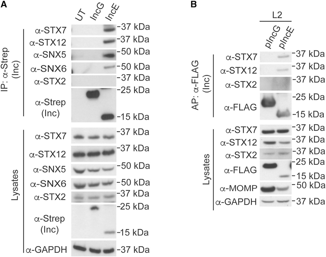
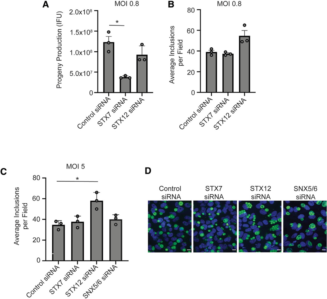
Chlamydia trachomatis, a leading cause of bacterial sexually transmitted infections, creates a specialized intracellular replicative niche by translocation and insertion of a diverse array of effectors (Incs [inclusion membrane proteins]) into the inclusion membrane. Here, we characterize IncE, a multifunctional Inc that encodes two non-overlapping short linear motifs (SLiMs) within its short cytosolic C terminus. The proximal SLiM, by mimicking just a small portion of an R-N-ethylmaleimide-sensitive factor adaptor protein receptor (SNARE) motif, binds and recruits syntaxin (STX)7- and STX12-containing vesicles to the inclusion. The distal SLiM mimics the sorting nexin (SNX)5 and SNX6 cargo binding site to recruit SNX6-containing vesicles to the inclusion. By simultaneously binding two distinct vesicle classes, IncE brings these vesicles in close apposition with each other at the inclusion to facilitate C. trachomatis intracellular development. Our work suggests that Incs may have evolved SLiMs to enable rapid evolution in a limited protein space to disrupt host cell processes.

Keywords: CP: Microbiology; Chlamydia trachomatis; SNARE; SNARE protein; host-pathogen interactions; inclusion membrane protein; intracellular bacteria; microbial pathogenesis; short linear motif; sorting nexin; syntaxin; vesicular trafficking.


Conflict of interest statement

Declaration of interests The authors declare no competing interests.

Figures

Figure 1.
Figure 1.. IncE binds specifically to STX7 and STX12
Co-APs of lysates from (A) HEK293T cells transfected with IncGStrep or IncEStrep or (B) HeLa cells infected with L2+pIncGFLAG or L2+pIncEFLAG in the presence of inducer. The unrelated STX2 serves as a control for binding specificity. GAPDH serves as a loading control. MOMP, a Ct protein, serves as an indicator of the efficiency of infection.
Figure 2.
Figure 2.. Defining the binding interface of STX7, STX12, and IncE
(A, C, and E) Schematic of (A) STX7, (C) STX12, and (E) IncE constructs. Habc, regulatory domain; TM, transmembrane. (B and D) Co-AP of transfected IncEStrep with transfected (B) GFP-STX7 or (D) GFP-STX12 deletion constructs. GAPDH serves as a loading control. (F) Co-AP of transfected IncEStrep constructs with endogenous STX7, STX12, or SNX2. IncE monomers and dimers are indicated by an asterisk. SNX2 serves as a loading control as well as a positive control for the ESCPE-1 complex (composed of SNX1/2/5/6). Data shown are representative of immunoblots from two biological replicates.
Figure 3.
Figure 3.. The IncE StxBM and SnxBM are non-overlapping
(A and B) Co-APs of lysates from HeLa cells infected with L2ΔincE expressing the indicated IncE constructs with (A) mCherry-SNX5, STX7, and STX12 or (B) GFP-SNX6, STX7, and STX12. Data shown are representative of immunoblots from three biological replicates. GAPDH serves as a loading control. MOMP serves as an indicator of the efficiency of infection.
Figure 4.
Figure 4.. IncE StxBM is required for late steps of infection
(A and B) Intra-strain comparisons of (A) inclusion formation at 24 hpi and (B) progeny production at 48 hpi. Infections were performed at a low MOI (MOI = 1) and compared to the indicated strain without an inducer. (C) Inter- and intra-strain quantification of inclusion fusion at 24 hpi. Infections were performed at a high MOI (MOI = 5). Data shown are averages ± SEM for at least three biological replicates (indicated with circle), with at least 99 fields counted for each replicate. *p < 0.05, **p < 0.02, and ***p < 0.0005; Welch’s ANOVA.
Figure 5.
Figure 5.. STX7 and STX12 serve distinct functions during infection
Control small interfering RNA (siRNA)-, STX7 siRNA-, or STX12 siRNA-depleted cells infected at a low MOI (~0.8, A and B) or high MOI (~5, C and D) for 24 h. Quantification of (A) progeny production and (B and C) average inclusions per field. (A and B) Shown is the average ± SEM for three technical replicates with a total of 33 fields. (C) Data shown are averages ± SEM for three biological replicates, with a total of 99 fields counted. The decreased inclusion fusion in the STX12 siRNA-treated cells is reflected by an increase in the average number of inclusions per field. (D) Representative single-slice confocal immunofluorescence images (scale bar: 10 μm). Multiple smaller unfused inclusions are present in the STX12 siRNA-treated sample. *p < 0.05 and **p < 0.005; Welch’s ANOVA.
Figure 6.
Figure 6.. IncE is required to tether distinct vesicle classes together at the inclusion
(A) Schematic and representative single-slice confocal immunofluorescence images depicting quantitation of STX7/STX12/SNX6 overlap in vesicles (insets) proximal and distal to inclusion. Fluorescence profiles on vesicles proximal (≤ 1 μm) and distal (>5 μm) to the inclusion were plotted for transfected GFP-STX7 (green), transfected mCh-STX12 (magenta), and endogenous SNX6 (blue). The maximum fluorescent intensity offsets of GFP-STX7 and mCh-STX12 from SNX6 were computed. Scale bar: 5 μm. (B and C) Quantification of STX7/SNX6 (green) and STX12/SNX6 (magenta) offset in (B) vesicles proximal or distal to inclusion in cells infected for 24 h with L2+vector or L2ΔincE+vector or (C) vesicles proximal to the inclusion in cells infected for 24 h with L2+vector, L2ΔincE+vector, or the indicated L2ΔincE+pIncE variants. All experiments were performed in the presence of an inducer. Shown are individual data points as well as the average ± SD for 30 vesicles for each infection condition. ***p < 0.0005; Welch’s ANOVA.
Figure 7.
Figure 7.. Model of IncE:STX7/STX12 and IncE:SNX5/SNX6 interactions
IncE encodes two SLiMs, the StxBM and SnxBM, which bind to STX7/12 and SNX5/6, respectively. The binding of the IncE SnxBM to the SNX5/6 cargo binding domain reroutes SNX6+, and presumably SNX5+ (gray half-moon), vesicles to the inclusion. By binding to SNX5/6, IncE displaces SNX5/6 cargo proteins and disrupts ESCPE-1-mediated restriction of Ct intracellular development. IncE StxBM binding to STX7 (tan squiggle line) and STX12 (tan squiggle line) reroutes STX7- and STX12-containing vesicles to the inclusion. At or near the inclusion, the STX7/12- and SNX5/6-containing vesicles are brought in close apposition in an IncE-dependent manner and may fuse with each other and/or with the inclusion membrane. STX7 contributes to the production of infectious progeny, while STX12 functions to promote efficient inclusion fusion.

Update of

References

    1. Hocking JS, Geisler WM, and Kong FYS (2023). Update on the Epidemiology, Screening, and Management of Chlamydia trachomatis Infection. Infect. Dis. Clin. North Am 37, 267–288. 10.1016/j.idc.2023.02.007. - DOI - PubMed
    1. Elwell C, Mirrashidi K, and Engel J (2016). Chlamydia cell biology and pathogenesis. Nat. Rev. Microbiol 14, 385–400. 10.1038/nrmicro.2016.30. - DOI - PMC - PubMed
    1. Rockey DD, Scidmore MA, Bannantine JP, and Brown WJ (2002). Proteins in the chlamydial inclusion membrane. Microbes Infect. 4, 333–340. - PubMed
    1. Mirrashidi KM, Elwell CA, Verschueren E, Johnson JR, Frando A, Von Dollen J, Rosenberg O, Gulbahce N, Jang G, Johnson T, et al. (2015). Global Mapping of the Inc-Human Interactome Reveals that Retromer Restricts Chlamydia Infection. Cell Host Microbe 18, 109–121. 10.1016/j.chom.2015.06.004. - DOI - PMC - PubMed
    1. Simonetti B, Paul B, Chaudhari K, Weeratunga S, Steinberg F, Gorla M, Heesom KJ, Bashaw GJ, Collins BM, and Cullen PJ (2019). Molecular identification of a BAR domain-containing coat complex for endosomal recycling of transmembrane proteins. Nat. Cell Biol 21, 1219–1233. 10.1038/s41556-019-0393-3. - DOI - PMC - PubMed

Publication types

LinkOut - more resources