. 2024 Dec 3;147(12):4043-4055.
doi: 10.1093/brain/awae266.

Exon 1-targeting miRNA reduces the pathogenic exon 1 HTT protein in Huntington's disease models

Affiliations

Exon 1-targeting miRNA reduces the pathogenic exon 1 HTT protein in Huntington's disease models

Marina Sogorb-Gonzalez et al. Brain. .

Abstract

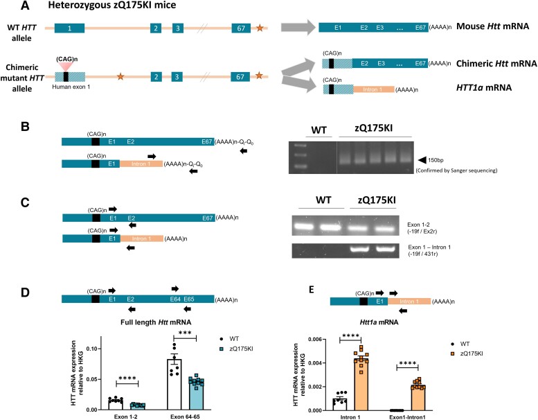
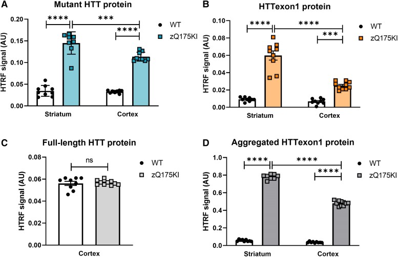
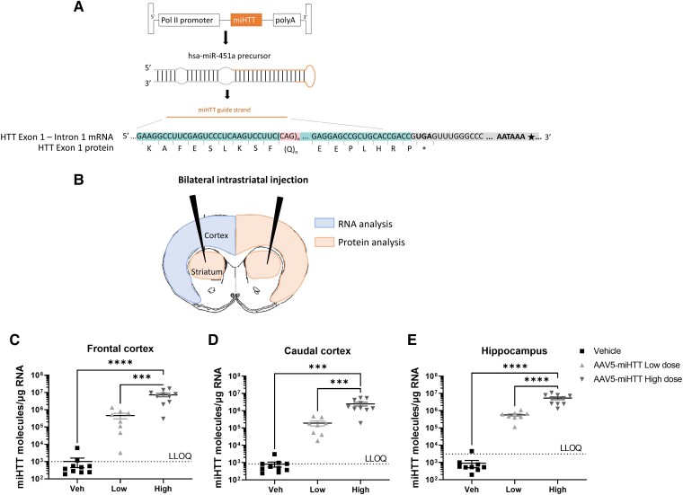
Huntington's disease (HD) is a fatal neurodegenerative disease caused by a trinucleotide repeat expansion in exon 1 of the huntingtin gene (HTT) that results in toxic gain of function and cell death. Despite its monogenic cause, the pathogenesis of HD is highly complex, and increasing evidence indicates that, in addition to the full-length (FL) mutant HTT protein, the expanded exon 1 HTT (HTTexon1) protein that is translated from the HTT1a transcript generated by aberrant splicing is prone to aggregate and might contribute to HD pathology. This finding suggests that reducing the expression of HTT1a might achieve a greater therapeutic benefit than targeting only FL mutant HTT. Conversely, strategies that exclusively target FL HTT might not completely prevent the pathogenesis of HD. We have developed an engineered microRNA targeting the HTT exon 1 sequence (miHTT), delivered via adeno-associated virus serotype 5 (AAV5). The target sequence of miHTT is present in both FL HTT and HTT1a transcripts. Preclinical studies with AAV5-miHTT have demonstrated efficacy in several rodent and large animal models by reducing FL HTT mRNA and protein and rescuing HD-like phenotypes and have been the rationale for phase I/II clinical studies now ongoing in the USA and Europe. In the present study, we evaluated the ability of AAV5-miHTT to reduce the levels of aberrantly spliced HTT1a mRNA and the HTTexon1 protein in the brain of two mouse models of HD (heterozygous zQ175 knock-in mice and humanized Hu128/21 mice). Polyadenylated HTT1a mRNA and HTTexon1 protein were detected in the striatum and cortex of heterozygous zQ175 knock-in mice, but not in wild-type littermate control mice. Intrastriatal administration of AAV5-miHTT resulted in dose-dependent expression of mature miHTT microRNA in cortical brain regions, accompanied by significant lowering of both FL HTT and HTT1a mRNA expression at 2 months postinjection. Mutant HTT and HTTexon1 protein levels were also significantly reduced in the striatum and cortex of heterozygous zQ175 knock-in mice at 2 months after AAV5-miHTT treatment and in humanized Hu128/21 mice 7 months post-treatment. The effects were confirmed in primary Hu128/21 neuronal cultures. These results demonstrate that AAV5-miHTT gene therapy is an effective approach to lower both FL HTT and the pathogenic HTTexon1 levels, which could potentially have an additive therapeutic benefit in comparison to other HTT-targeting modalities.

Keywords: HTT exon 1; AAV5-miHTT; Huntington’s disease; Huntington’s disease mouse models; aberrant splicing; gene therapy.


Conflict of interest statement

A.S., A.V. and M.E. are employees of uniQure and may own stock and/or stock options. Filed patent applications pertaining to the results presented in this paper include the following: RNA interference induced HTT gene suppression (WO2016/102664, resulting in at least US 10,174,321, US 10,767,180 and EP 3237618B1), A companion diagnostic to monitor the effects of gene therapy (PCT/EP2019/081759), Method and means to deliver miRNA to target cells (PCT/EP2019/081822) and Targeting misspliced transcripts in genetic disorders (PCT/EP2020/075871); the last three have not yet been published.

Figures

Figure 1
Figure 1
Detection of the Htt1a transcript in the cortex of zQ175 knock-in mice. (A) Schematic representation of wild-type (WT) huntingtin (Htt) allele and chimeric mutant Htt allele in heterozygous zQ175 knock-in (KI) mice. (B) 3′RACE RT-PCR together with intron 1 Htt primers showed the presence of polyadenylated Htt1a mRNA in zQ175 KI brain cortex, but not in WT mice. (C) Detection of polyadenylated HTT1a mRNA in the cortex of zQ175 KI mice by oligo-dT RT-PCR assay. Spliced exon 1–exon 2 transcripts (top) were detected in frontal cortex of both WT and zQ175 KI mice, whereas exon 1–intron 1 product (bottom) was detected only in zQ175 KI mice, but not in WT. (D) Relative expression of FL-Htt in the frontal cortex of zQ175 KI mice and WT detected by TaqMan qPCR with primer-probe sets exon1–2 (unpaired t-test, ****P < 0.001) and exon64–65 (unpaired t-test, ***P = 0.003). (E) Relative expression of Htt1a mRNA in cortical tissue of zQ175 KI and WT mice detected by TaqMan qPCR with primer–probe sets intron 1 (unpaired t-test, ****P < 0.0001) and human exon 1–intron 1 (unpaired t-test, ****P < 0.0001). In D and E, bars show the mean ± standard error of the mean (SEM).
Figure 2
Figure 2
Detection of HTTexon1 protein and other huntingtin (HTT) protein species in zQ175 knock-in mice. (A) Levels of soluble mutant HTT protein (FL-HTT and HTTexon1) in striatum and frontal cortex of wild-type (WT) and zQ175 knock-in (KI) mice measured by homogeneous time-resolved fluorescence (HTRF) with 2B7 and 4C9 antibodies and represented as the mean ± SEM (one-way ANOVA, Tukey’s multiple comparisons test, ***P = 0.009, ****P < 0.001). (B) Levels of soluble HTTexon1 protein in striatum and frontal cortex of WT and zQ175 KI mice measured by HTRF with 2B7 and MW8 antibodies and represented as the mean ± SEM (one-way ANOVA, Tukey’s multiple comparisons test, ****P < 0.001). (C) Levels of soluble full-length HTT protein (mutant and WT) in frontal cortex of WT and zQ175 KI mice measured by HTRF with MAB5490 and MAB2166 antibodies and represented as the mean ± SEM (unpaired t-test, ns, P = 0.7613). (D) Levels of aggregated HTTexon1 protein in striatum and frontal cortex of WT and zQ175 KI mice measured by HTRF with 4C9 and MW8 antibodies and represented as the mean ± SEM (one-way ANOVA, Tukey’s multiple comparisons test, ****P < 0.001).
Figure 3
Figure 3
Intrastriatal administration of AAV5-miHTT results in dose-dependent expression of HTT exon 1-targeting miHTT. (A) Schematic representation of AAV5-delivered expression cassette including Pol II promoter, exon 1-targeting miHTT transgene and polyA signal. The transgene is processed into pre-miRNA hairpin with the same structure as has-miR-451 precursor, then processed into an miHTT guide strand that is complementary to HTT exon 1 sequence upstream CAG repeat expansion. (B) Representation of bilateral intrastriatal injection and collection of colour-coded brain tissues for RNA and protein analysis. (CE) miHTT transgene expression in frontal cortex (C), caudal cortex (D) and hippocampus (E) from the left hemisphere determined by custom TaqMan RT-qPCR and represented as miHTT molecules per microgram of RNA (mean ± SEM) per dose group (n = 8–10, one-way ANOVA, Tukey’s multiple comparisons test, ***P < 0.005, ****P < 0.001).
Figure 4
Figure 4
AAV5-miHTT shows dose-dependent lowering of full-length Htt and Htt1a mRNA and HTT and HTTexon1 protein in zQ175 knock-in mice at 2 months postinjection. (A) Expression level of FL-Htt mRNA in right frontal cortex of AAV5-miHTT-treated mice relative to vehicle group. (B) Expression level of Htt1a mRNA in right frontal cortex of AAV5-miHTT-treated mice relative to vehicle group. (C) Levels of soluble mutant HTT protein in striatum and cortex of AAV5-miHTT-treated mice relative to vehicle group and wild-type (WT) mice. (D) Levels of HTTexon1 HTT protein in striatum and cortex of AAV5-miHTT-treated mice relative to vehicle group and WT mice. (E) Levels of aggregated HTTexon1 HTT protein in striatum and cortex of AAV5-miHTT-treated mice relative to vehicle group and WT mice. (F) Levels of full-length HTT (mutant and WT) protein in frontal cortex of AAV5-miHTT-treated mice relative to vehicle group. In AF, bars represent the mean ± SEM. Statistics: one-way ANOVA, Tukey’s multiple comparisons test (*P < 0.05, **P < 0.005, ***P < 0.0005 and ****P < 0.0001).
Figure 5
Figure 5
Striatal AAV5-miHTT treatment lowers levels of different huntingtin (HTT) protein species, including HTTexon1, in the hippocampus of Hu128/21 mice. (A) Four different HTT homogeneous time-resolved fluorescence (HTRF) assays were used, measuring soluble mutant HTT protein (2B7–MW1), soluble HTTexon1 protein (2B7–MW8), full-length HTT protein (MAB5490–MAB2166) and aggregated HTTexon1 (4C9–MW8), all of them demonstrating dose-dependent lowering after AAV5-miHTT treatment in Hu128/21 hippocampi (data are represented as a percentage of Veh-treated groups). (BE) Data from the four different HTT HTRF assays in Hu21/21 and Hu128/21, expressed in arbitrary units (AU), with dose-dependent lowering in Hu128/21 but not in Hu21/21 mice, in which the analytes were detected only at background levels. In AE, bars represent the mean ± SEM. Statistics: one-way ANOVA, Tukey’s multiple comparisons test (*P < 0.05, **P < 0.005, ***P < 0.0005 and ****P < 0.0001).
Figure 6
Figure 6
Soluble huntingtin (HTT) protein expression in primary Hu128/21 neurons treated with AAV5-GFP or AAV5-miHTT. Cells were transduced with AAV5-miHTT at a multiplicity of infection of 105, 106 or 107. Control groups included vehicle-treated and AAV5-GFP multiplicity of infection 107-treated cells. Five days post-transduction, cell pellets were collected to determine soluble HTT protein levels, analysed by homogeneous time-resolved fluorescence (HTRF) using different antibody pairs. Two independent experiments were performed, with three biological replicates per condition. HTT expression levels (biological replicates and mean ± SEM) are represented as a percentage of vehicle-treated control groups: (A) total soluble mutant HTT (FL-HTT and HTTexon1; MW1–4C9 antibody pair); (B) soluble HTTexon1 (2B7–MW8 antibody pair); and total soluble full-length HTT (mutant and WT) with (C) MAB5490–MAB2166 antibody pair or (D) D7F7–MAB5490 antibody pair. Each experiment was evaluated independently by one-way ANOVA followed by multiple comparison tests, corrected using Sidak’s test (*P < 0.05, **P < 0.01, ***P < 0.001 and ****P < 0.0001 versus GFP controls).

References

    1. MacDonald ME, Ambrose CM, Duyao MP, et al. . A novel gene containing a trinucleotide repeat that is expanded and unstable on Huntington’s disease chromosomes. Cell. 1993;72:971–983. - PubMed
    1. Tabrizi SJ, Schobel S, Gantman EC, et al. . A biological classification of Huntington’s disease: The integrated staging system. Lancet Neurol. 2022;21:632–644. - PubMed
    1. Stoyas CA, La Spada AR. The CAG–polyglutamine repeat diseases: A clinical, molecular, genetic, and pathophysiologic nosology. In: Geschwind DH, Paulson HL, eds. Handbook of clinical neurology. Vol. 147. Elsevier; 2018:143–170. - PubMed
    1. Takahashi T, Katada S, Onodera O. Polyglutamine diseases: Where does toxicity come from? What is toxicity? Where are we going? J Mol Cell Biol. 2010;2:180–191. - PubMed
    1. Arrasate M, Finkbeiner S. Protein aggregates in Huntington’s disease. Exp Neurol. 2012;238:1–11. - PMC - PubMed

Grants and funding