. 2024 Dec;49(13):2060-2068.
doi: 10.1038/s41386-024-01963-7. Epub 2024 Aug 18.

Basal forebrain-lateral habenula inputs and control of impulsive behavior

Affiliations

Basal forebrain-lateral habenula inputs and control of impulsive behavior

Eun-Kyung Hwang et al. Neuropsychopharmacology. 2024 Dec.

Abstract

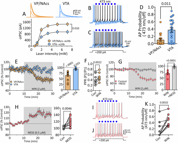
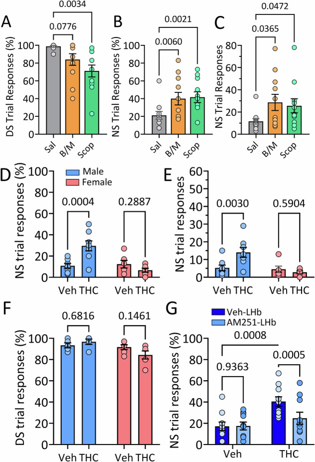
Deficits in impulse control are observed in several neurocognitive disorders, including attention deficit hyperactivity (ADHD), substance use disorders (SUDs), and those following traumatic brain injury (TBI). Understanding brain circuits and mechanisms contributing to impulsive behavior may aid in identifying therapeutic interventions. We previously reported that intact lateral habenula (LHb) function is necessary to limit impulsivity defined by impaired response inhibition in rats. Here, we examine the involvement of a synaptic input to the LHb on response inhibition using cellular, circuit, and behavioral approaches. Retrograde fluorogold tracing identified basal forebrain (BF) inputs to LHb, primarily arising from ventral pallidum and nucleus accumbens shell (VP/NAcs). Glutamic acid decarboxylase and cannabinoid CB1 receptor (CB1R) mRNAs colocalized with fluorogold, suggesting a cannabinoid modulated GABAergic pathway. Optogenetic activation of these axons strongly inhibited LHb neuron action potentials and GABA release was tonically suppressed by an endogenous cannabinoid in vitro. Behavioral experiments showed that response inhibition during signaled reward omission was impaired when VP/NAcs inputs to LHb were optogenetically stimulated, whereas inhibition of this pathway did not alter LHb control of impulsivity. Systemic injection with the psychotropic phytocannabinoid, Δ9-tetrahydrocannabinol (Δ9-THC), also increased impulsivity in male, and not female rats, and this was blocked by LHb CB1R antagonism. However, as optogenetic VP/NAcs pathway inhibition did not alter impulse control, we conclude that the pro-impulsive effects of Δ9-THC likely do not occur via inhibition of this afferent. These results identify an inhibitory LHb afferent that is controlled by CB1Rs that can regulate impulsive behavior.


Conflict of interest statement

The authors declare no competing interests.

Figures

Fig. 1
Fig. 1. Basal forebrain (BF) neurons located in the nucleus accumbens shell (NAcs) and the ventral pallidum (VP) project to the LHb and express CB1R mRNA, GADs mRNA, or both.
A Retrograde tracer FG was delivered into the LHb. B FG-IR in the injection site (brown). C Low magnification of a NAc shell (d) and VP (e) coronal section showing detection of FG-immunoreactivity (FG-IR, blue), expression of CB1 mRNA (red), and expression of GADs mRNA (green). These areas in yellow boxes are shown at higher magnification in the sequence shown in (E), top and bottom, respectively. D Number of FG-IR cells detected in VP and NAcs, sorted by co-expression of CB1R mRNA and/or GADs mRNA. A total of 208 FG-IR VP neurons projecting to the LHb were detected, and of these 24 (13%) expressed only CB1R mRNA, 59 (28%) expressed only GADs mRNA, 80 (38%) co-expressed CB1R and GADs mRNA, and 45 (21%) lacked both CB1R and GADs mRNA. A total of 369 FG-IR NAcs neurons projecting to the LHb were detected, and of these 97 (26%) expressed only CB1 mRNA, 79 (21%) expressed only GADs mRNA, 67 (18%) co-expressed CB1R and GADs mRNA, and 126 (34%) lacked both CB1R and GADs mRNA. FG-IR cell counts were made between +1.92 and +0.72 mm from bregma (n = 3 rats, 8–9 sections per rat). E NAcs and VP areas corresponding to boxes (d, e) in panel (C) at higher magnification. FG-IR neurons expressing CB1R mRNA without GADs mRNA are indicated by single arrows, FG-IR neurons expressing GADs mRNA and CB1R mRNA are indicated by single arrow heads, and double arrow heads indicate FG-IR without GADs or CB1R mRNA in NAcs. A FG-IR neuron expressing GADs mRNAs, without CB1R mRNA in VP is indicated by double arrows. Abbreviations; aca, anterior commissure; LHb, lateral habenula; MHb, medial habenula; fr, fasciculus retroflexus.
Fig. 2
Fig. 2. Synaptic currents evoked by ChR2 stimulation in LHb after transfection of VP/NAcs or VTA in are primarily mediated by GABA and not glutamate or glycine.
A Diagram showing sites of ChR2 construct injection into VP/NAcs and photomicrographs showing eYFP fluorescence at the injection site and in the habenula ~8 weeks after injection. B Diagram showing injection sites and eYFP after VTA injections of ChR2 construct. Number at left indicate sections relative to bregma. Mean photostimulation-evoked current sweeps obtained during control periods, and during sequential application of TTX, 4-AP, and PTX in LHb neurons from rats transfected with AAV-ChR2 in the VP/NAcs (C) or VTA (D). The representative mean time courses for these experiments are shown in (E, F). In (G), the 10–90% rise times are shown for both inputs to LHb, and (H) shows the time constant for the decay of the synaptic currents (tau) evoked by each LHb input. These kinetic measures were similar for both pathways (unpaired t test; rise time, t13 = 0.79, p = 0.45, decay time constant, t13 = 0.59, p = 0.73, respectively). I Mean time course for synaptic currents evoked via photostimulation at each pathway during application of the AMPAR antagonist DNQX (10 µM) and glycine receptor antagonist strychnine (5 µM) at a holding potential of 0 mV. J Mean current-voltage (I-V) relationships for photoactivated synaptic currents from VP/NAcs and VTA inputs to LHb neurons. The calculated reversal potential for Cl- is indicated by downward arrow. Above are signal averaged synaptic currents collected during activation of each pathway across a range of membrane holding potentials (Vm). The synaptic current I-V curves reversed near that predicted for Cl- ions (ECl-, see text). Number of cells/rats: (E), 5/3; (F), 6/4; (G–I), 10/16; (J), VTA, 2/3; VP/NAcs, 4/7.
Fig. 3
Fig. 3. Relative strength of inhibition of LHb neurons by VTA and VP/NAcs afferents and sensitivity to cannabinoids in vitro.
A Relationship between 473 nm laser power (single -pulses, 2 ms duration) and oIPSC amplitude in LHb neurons from wildtype rats injected with AAV-ChR2 in either VTA or VP/NAcs. VP/NAcs inputs to LHb generate significantly larger oIPSCs than those from VTA. B, C Action potentials (AP) generated by injection of +200 pA (1 s) currents in LHb neurons. Waveforms in gray were recorded without activation of VP/NAcs or VTA inputs, whereas AP wave forms shown in blue were recorded during stimulation of ChR2 by 473 nm light (blue circles) at VP/NAcs (B) or VTA (C) inputs (1 s photostimulation train = seven-2 msec duration pulses delivered at 7 Hz at 145 ms intervals during LHb neuron depolarization). D Summary of effect of activation of VP/NAcs (n = 18) or VTA (n = 24) LHb input by ChR2 on the probability of AP discharge. Activation of either input significantly reduced AP probability, but the VP/NAcs input was significantly more effective at silencing LHb neurons t40 = 3.526, p = 0.011, (unpaired t test). E Effect of bath application of the cannabinoid agonist WIN55212-2 (WIN, 2 µM) on oIPSCs evoked by ChR2 at VP/NAcs (n = 18 cells) or VTA (n = 8 cells) inputs to LHb neurons. Whereas WIN had a small effect on IPSCs from VTA input, the effect on VP/NAcs input was significantly larger (p = 0.0001, t test). The Y-axis title for bar graph is the same as time course. (F). WIN significantly increased the paired-oIPSC ratio (PPR) at VP/NAcs inputs to LHb, indicating a presynaptic effect (paired t test, t7 = 4.7, p = 0.022). G Preincubation of LHb cells with the neutral CB1R antagonist NESS 0327 (NESS) prevents the inhibition of VP/NAcs oIPSCs in LHb (one-sample t test, WIN = t7 = 6.76, p = 0.0003; WIN + NESS = t7 = 1.16, p = 0.284). Left and right graph share y-axis. H CB1R antagonism by NESS reveals tonic eCB suppression of VP/NAcs oIPSCs in LHb neurons. Left and right panels share y-axis label. I AP waveforms generated by current injection with ChR2-activation of VP/NAcs inputs (red) or without ChR2-activation (gray) in control aCSF. J AP waveforms recorded with and without ChR2-activation of VP/NAcs inputs to LHb during WIN application. K AP probability before (gray circles) and during application of WIN (red circles). The reduction of synaptic inhibition by WIN significantly increased AP probability (paired t test, t9 = 4.76, p = 0.001). Number of cells/rats: A VP/NAcs to LHb: 8/5, VTA to LHb, 8/5; (D) VP/NAcs to LHb, 18/13, VTA to LHb, 24/16; (E) VP/NAcs, 8/6, VTA, 8/5; (F). 8/5; (G). WIN alone, 8/5: WIN + NESS, 8/5; (H) 11/8; (K) 10/7.
Fig. 4
Fig. 4. Effects of pharmacological manipulation of the LHb on response inhibition in wildtype rats.
A–C Effects of intra-LHb infusion of Saline (Sal), baclofen/muscimol (B/M) or scopolamine (Scop) on DS-NS responding (n = 8 male and 3 female rats). A Scop infusion into LHb significantly decreased responding during trials in which food pellet availability was signaled (DS, repeated measures 1-way ANOVA, F1.64,16.4 = 6.95, p = 0.009, p values from Dunnett’s post hoc test). B Scop and B/M significantly increased the percentage of NS trials (when reward was not available) in which responses occurred (repeated measures 1-way ANOVA, F1.4,14 = 7.79, p = 0.009, p values from Dunnett’s post hoc test). C Scop and B/M significantly increased the number of NS trial responses (repeated measures 1-way ANOVA, F1.67,16.73 = 3.87, p = 0.0478, p values from Dunnett’s post hoc test). D–G Effects of systemic injection of Δ9-THC (1 mg/kg) or vehicle on DS-NS responding in male and female rats. D Δ9-THC injection increased the percentage of NS trials in which responses were observed in male rats only (2-way mixed effects ANOVA, n = 8 males and 6 females, drug x sex interaction, F1,10 = 12.57, p = 0.0053). The numbers above bars in (D–G) represent post hoc comparison p values using the uncorrected Fisher’s Least Significant Difference test (uFLSD). The legend in (D) applies to panels (D–F). E Systemic Δ9-THC effect on the number of responses during NS trials in male and female rats (2-way ANOVA, Drug x Sex Interaction, F1,24 = 6.50, p = 0.0165). F No effect of Δ9-THC on percent of trials responding when reward availability was signaled (DS; 2-way mixed effects ANOVA, n = 10, F1,18 = 3.54, p = 0.080). G The increase in proportion of NS trial responses caused by systemic injection of Δ9-THC in male rats was prevented by infusion of AM251 into the LHb (n = 10; 2-Way RM ANOVA, systemic THC x AM251 infusion Interaction = F1,18 = 9.35, p = 0.0068).
Fig. 5
Fig. 5. Impaired response inhibition during activation of GABAergic BF input to the LHb by ChR2 in GADCre rats.
A Comparison of the percent of neutral stimulus (NS) trials in which responses on the active lever were observed in the absence (off) and presence of 455 nm light delivered via optical fibers terminating above the LHb, ~8 weeks after expression of Cre-dependent eYFP in the VP/NAcs (n = 6 rats; paired t test, t5 = 1.67, p = 0.155). B Optogenetic inhibition of VP/NAcs GABAergic inputs to LHb by stimulation of NpHR with 545 nm light does not affect NS responding (n = 4 rats; paired t test, t3 = 0.38, p = 0.728). C Activation of VP/NAcs GABAergic inputs by light stimulation of ChR2 in LHb significantly increased the percent of NS trials in which responses were observed (paired t test; n = 10 male rats, t9 = 3.44, p = 0.007). The total number of NS responses shown in Fig. S4A. D ChR2 photostimulation with 455 nm light significantly increases responding during presentation of the NS, and this is not significantly different between male and female GADCre rats (n = 16 male and 11 female rats; 2-way RM ANOVA, light x sex interaction, F1,25 = 1.546, p = 0.225; main effect of light, F1,25 = 41.03, p < 0.0001; Post hoc comparisons by uFLSD, effect of light in males, p < 0.0001; effect of light in females, p = 0.0025). ns, non-significant.

References

    1. Vonder Haar C, Lam FC, Adams WK, Riparip LK, Kaur S, Muthukrishna M, et al. Frontal traumatic brain injury in rats causes long-lasting impairments in impulse control that are differentially sensitive to pharmacotherapeutics and associated with chronic neuroinflammation. ACS Chem Neurosci. 2016;7:1531–42. - PMC - PubMed
    1. Flerlage WJ, Langlois LD, Rusnak M, Simmons SC, Gouty S, Armstrong RC, et al. Involvement of lateral habenula dysfunction in repetitive mild traumatic brain injury-induced motivational deficits. J Neurotrauma. 2023;40:125–40. - PMC - PubMed
    1. Beauchaine TP, Zisner AR, Sauder CL. Trait impulsivity and the externalizing spectrum. Annu Rev Clin Psychol. 2017;13:343–68. - PubMed
    1. Dougherty DM, Mathias CW, Marsh DM, Moeller FG, Swann AC. Suicidal behaviors and drug abuse: impulsivity and its assessment. Drug Alcohol Depend. 2004;76:S93–s105. - PubMed
    1. Ersche KD, Turton AJ, Pradhan S, Bullmore ET, Robbins TW. Drug addiction endophenotypes: impulsive versus sensation-seeking personality traits. Biol Psychiatry. 2010;68:770–3. - PMC - PubMed

MeSH terms

Substances

LinkOut - more resources