

Association between dichotomized VASARI feature and overall survival in glioblastoma patients: a single-institution propensity score matching analysis
- PMID: 39155364
- PMCID: PMC11330608
- DOI: 10.1186/s40644-024-00754-z
Association between dichotomized VASARI feature and overall survival in glioblastoma patients: a single-institution propensity score matching analysis
Abstract
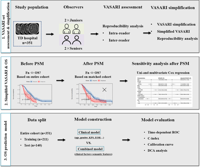
Objectives: This study aimed to investigate the intra- and inter-observer consistency of the Visually Accessible Rembrandt Images (VASARI) feature set before and after dichotomization, and the association between dichotomous VASARI features and the overall survival (OS) in glioblastoma (GBM) patients.
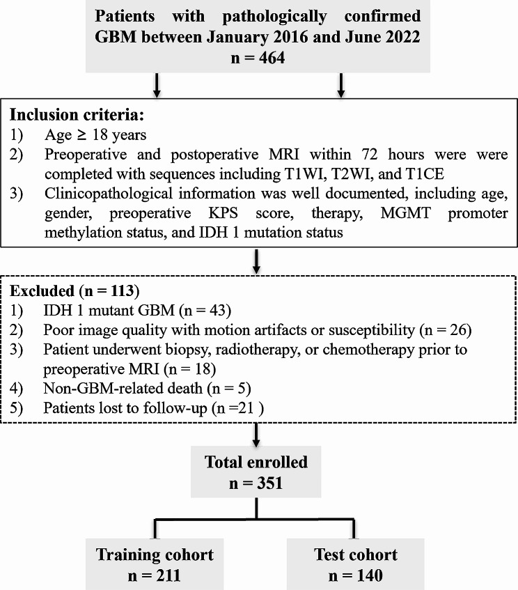
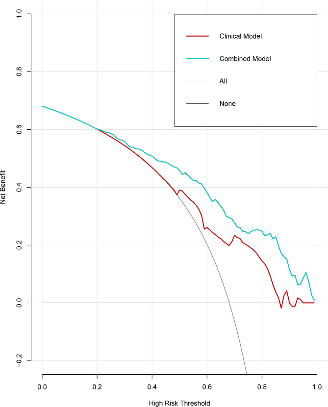
Methods: This retrospective study included 351 patients with pathologically confirmed IDH1 wild-type GBM between January 2016 and June 2022. Firstly, VASARI features were assessed by four radiologists with varying levels of experience before and after dichotomization. Cohen's kappa coefficient (κ) was calculated to measure the intra- and inter-observer consistency. Then, after adjustment for confounders using propensity score matching, Kaplan-Meier curves were used to compare OS differences for each dichotomous VASARI feature. Next, patients were randomly stratified into a training set (n = 211) and a test set (n = 140) in a 3:2 ratio. Based on the training set, Cox proportional hazards regression analysis was adopted to develop combined and clinical models to predict OS, and the performance of the models was evaluated with the test set.
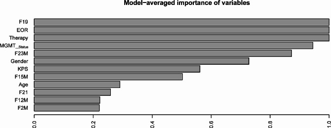
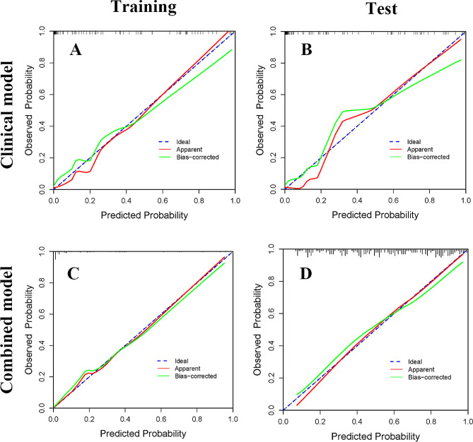
Results: Eleven VASARI features with κ value of 0.61-0.8 demonstrated almost perfect agreement after dichotomization, with the range of κ values across all readers being 0.874-1.000. Seven VASARI features were correlated with GBM patient OS. For OS prediction, the combined model outperformed the clinical model in both training set (C-index, 0.762 vs. 0.723) and test set (C-index, 0.812 vs. 0.702).
Conclusion: The dichotomous VASARI features exhibited excellent inter- and intra-observer consistency. The combined model outperformed the clinical model for OS prediction.
Keywords: Glioblastoma; Magnetic resonance imaging; Overall survival; Visually accessible rembrandt images.
© 2024. The Author(s).
Conflict of interest statement
The authors declare no competing interests.
The authors declare no conflicts of interest.
Figures
References
MeSH terms
Grants and funding
LinkOut - more resources
Full Text Sources
Medical
Miscellaneous
