. 2024 Aug 18;55(1):103.
doi: 10.1186/s13567-024-01358-y.

PRRSV hijacks DDX3X protein and induces ferroptosis to facilitate viral replication

Affiliations

PRRSV hijacks DDX3X protein and induces ferroptosis to facilitate viral replication

Qian Mao et al. Vet Res. .

Abstract

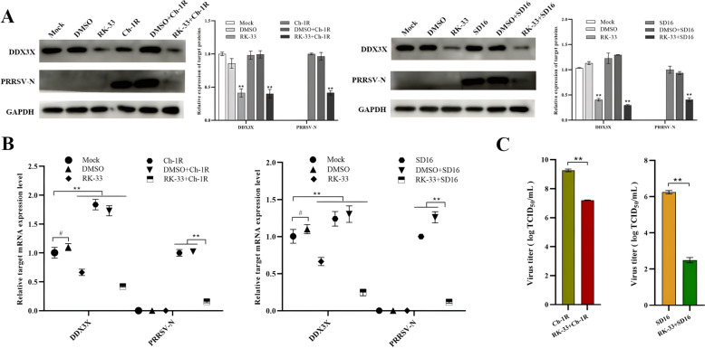
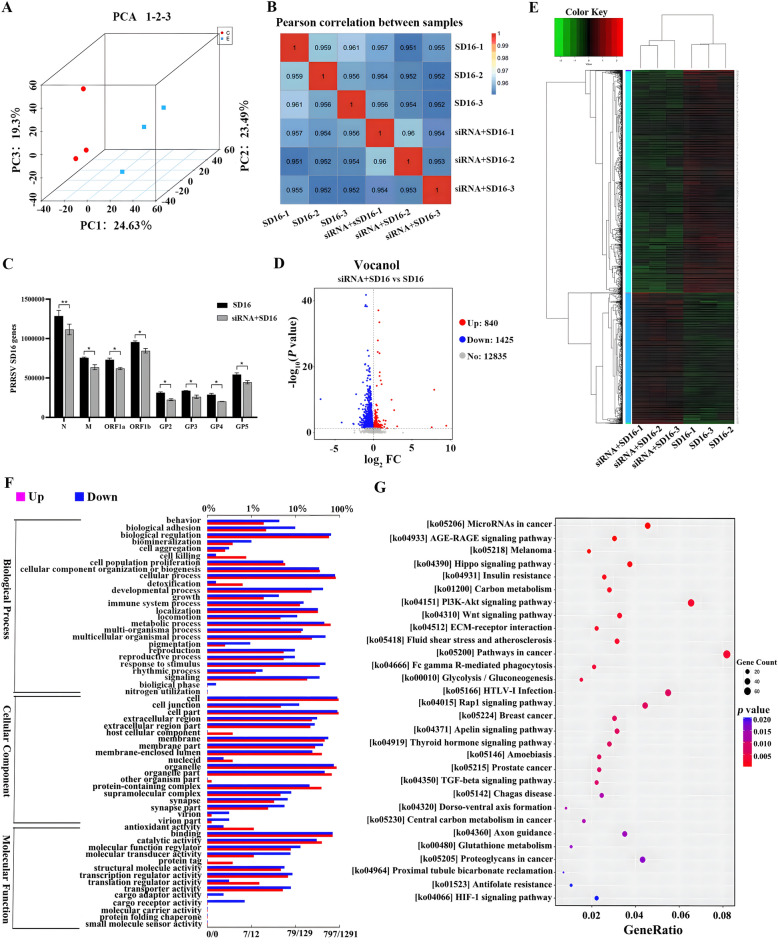
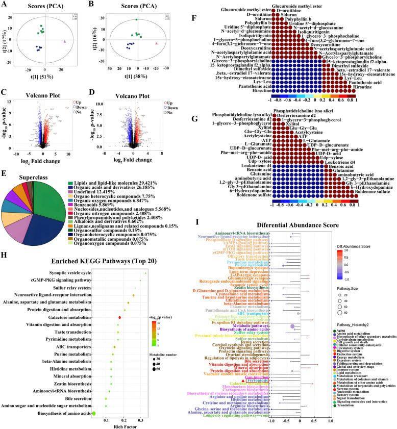
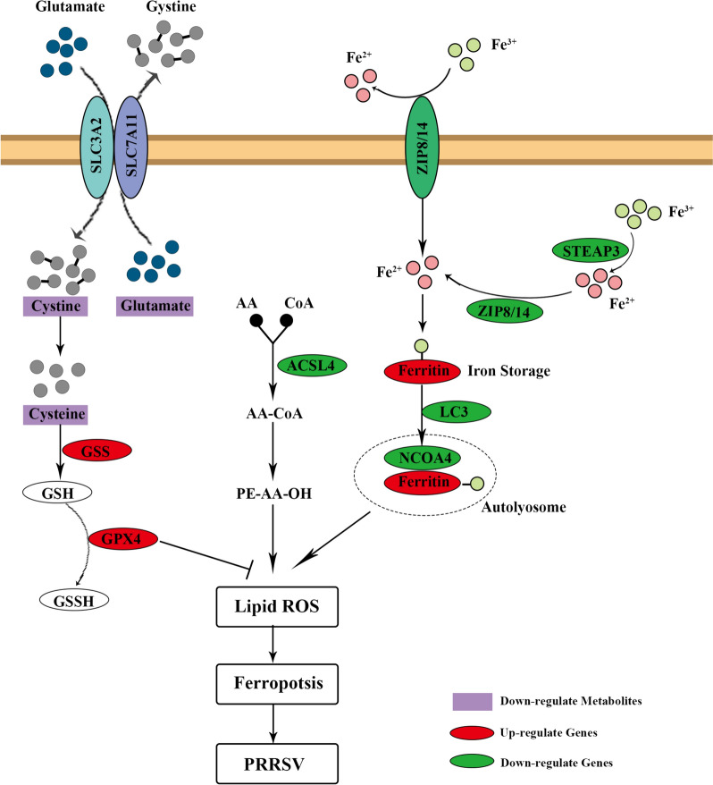
Porcine reproductive and respiratory syndrome virus (PRRSV) is a severe disease with substantial economic consequences for the swine industry. The DEAD-box helicase 3 (DDX3X) is an RNA helicase that plays a crucial role in regulating RNA metabolism, immunological response, and even RNA virus infection. However, it is unclear whether it contributes to PRRSV infection. Recent studies have found that the expression of DDX3X considerably increases in Marc-145 cells when infected with live PRRSV strains Ch-1R and SD16; however, it was observed that inactivated viruses did not lead to any changes. By using the RK-33 inhibitor or DDX3X-specific siRNAs to reduce DDX3X expression, there was a significant decrease in the production of PRRSV progenies. In contrast, the overexpression of DDX3X in host cells substantially increased the proliferation of PRRSV. A combination of transcriptomics and metabolomics investigations revealed that in PRRSV-infected cells, DDX3X gene silencing severely affected biological processes such as ferroptosis, the FoxO signalling pathway, and glutathione metabolism. The subsequent transmission electron microscopy (TEM) imaging displayed the typical ferroptosis features in PRRSV-infected cells, such as mitochondrial shrinkage, reduction or disappearance of mitochondrial cristae, and cytoplasmic membrane rupture. Conversely, the mitochondrial morphology was unchanged in DDX3X-inhibited cells. Furthermore, silencing of the DDX3X gene changed the expression of ferroptosis-related genes and inhibited the virus proliferation, while the drug-induced ferroptosis inversely promoted PRRSV replication. In summary, these results present an updated perspective of how PRRSV infection uses DDX3X for self-replication, potentially leading to ferroptosis via various mechanisms that promote PRRSV replication.

Keywords: DDX3X; PRRSV; ferroptosis; metabolome; transcriptome.


Conflict of interest statement

The authors declare that they have no competing interests.

Figures

Figure 1
Figure 1
mRNA and protein expression levels of DDX3X in PRRSV-infected Marc-145 cells. A, B Western blot and RT-qPCR analysis of the expression of DDX3X in PRRSV-infected Marc-145 cells determined at different time points post-infection. C, D Western blot and RT-qPCR analysis of the expression of DDX3X in Marc-145 cells infected with varying doses of PRRSV. E, F Western blot and RT-qPCR analysis of the expression of DDX3X in Marc-145 cells infected with live or inactivated viruses. Each of the experiments was independently repeated in triplicate. Two-tailed Student’s t-test determined statistical analysis of comparisons, n = 3 ± SEM. The stars indicate significant differences (*P < 0.05 or **P < 0.001).
Figure 2
Figure 2
Expression of DDX3X and virus proliferation in RK-33-treated and virus-infected Marc-145 cells. A, B Western blot and RT-qPCR analysis of the expressions of DDX3X and PRRSV-N protein. C Virus titres of PRRS viruses in RK-33-treated and virus-infected Marc-145 cells were detected at 48 hpi. Two-tailed Student’s t-test determined statistical analysis of comparisons, n = 3 ± SEM. The stars indicate significant differences (*P < 0.05 or **P < 0.001).
Figure 3
Figure 3
Expression of DDX3X and virus replication in Marc-145 cells with distinct RK-33 treatments. A Schematic of the time-of-addition experiment of RK-33 treatments. The Marc-145 cells were infected with PRRSV Ch-1R or SD16 at an MOI of 0.1 before being treated with RK-33: pre (− 12–0 h), during (0–1 h) and post (1–48 h) virus infection. DMSO was added at the same time as a mock control. B, C DDX3X and PRRSV-N protein expressions in Marc-145 cells treated with RK-33 at different time points and infected with PRRSV were determined at 48 hpi by western blotting and RT-qPCR. D Virus titres of PRRS viruses in Marc-145 cells treated with RK-33 were detected at 48 hpi. Two-tailed Student’s t-test determined statistical analysis of comparisons, n = 3 ± SEM. The stars indicate significant differences (*P < 0.05 or **P < 0.001).
Figure 4
Figure 4
Gene expression and virus proliferation in Marc-145 cells transfected with DDX3X-specific siRNA. A, B DDX3X and PRRSV-N protein expressions in Marc-145 cells transfected with DDX3X-specific siRNA were analysed by western blot and RT-qPCR at 48 hpi. C Virus titres of PRRS viruses in Marc-145 cells transfected with DDX3X-specific siRNA. Two-tailed Student’s t-test determined statistical analysis of comparisons, n = 3 ± SEM. The stars indicate significant differences (*P < 0.05 or **P < 0.001).
Figure 5
Figure 5
Gene expression and virus proliferation in Marc-145 cells transfected with pEGFP-N1-DDX3X plasmid. A, B Cells transfected with pEGFP-N1-DDX3X or empty pEGFP-N1 were infected with PRRSV before western blotting and RT-qPCR analysis. C Marc-145 cells were infected with PRRSV Ch-1R or SD16 12-h post-transfection with pEGFP-N1-DDX3X or empty pEGFP-N1. Supernatants were then collected for TCID50 measurement. Two-tailed Student’s t-test determined statistical analysis of comparisons, n = 3 ± SEM. The stars indicate significant differences (*P < 0.05 or **P < 0.001).
Figure 6
Figure 6
Transcriptomics analysis of Marc-145 cells transfected with DDX3X-specific siRNA and infected with PRRSV. A PCA plot of RNA-seq data. B Heatmap of correlation between samples from different groups. C Transcript levels of PRRSV N, M, ORF1a, ORF1b, GP2, GP3, GP4 and GP5 genes. D Volcanic map of the DEGs. E Heatmap of the DEGs. F GO terms of the DEGs. The lower x-axis represents the number of genes annotated to GO terms, while the upper x-axis represents the proportion of the total number of genes. Red lines represent up-regulated genes, and blue lines represent down-regulated ones. G KEGG enrichment bubble plot. The abscissa was the rate of the number of DEGs to the total number of DEGs, with the ordinate being the KEGG pathway. Two-tailed Student’s t-test determined statistical analysis of comparisons, n = 3 ± SEM. The stars indicate significant differences (*P < 0.05 or **P < 0.001).
Figure 7
Figure 7
Metabolomics of the PRRSV-infected Marc-145 cells transfected with DDX3X-specific siRNA. A, B PCA plot in positive ion mode (left) and negative ion mode (right). C, D Volcanic diagram of DEMs in positive ion mode (left) and negative ion mode (right). E Percentage of DEMs by chemical classification. F, G Heatmap of correlations in positive ion mode (left) or negative ion mode (right). H Top 20 KEGG enrichment pathway bubble maps. The colour from green to red indicates the successively decreased p-values. I Differential abundance scores of the KEGG pathway are classified according to the hierarchy classification method.
Figure 8
Figure 8
The combinate analysis of metabolomics and transcriptomics. A, B Venn diagrams showing the DMGs and DEMs of positive (left) or negative (right) ion modes in DDX3X-silenced cells compared with that in SD16-infected mock cells. C, D The top 20 pathways with the highest number of genes and metabolites in positive (left) or negative (right) ion modes. E, F Bubble diagram of the KEGG pathway enrichment analysis.
Figure 9
Figure 9
PRRSV-induced ferroptosis in DDX3X-silenced Marc-145 cells. A Electron microscopy image of mitochondria in Marc-145 cells. B Relative expression levels of ferroptosis-related genes GPX4, ACST4, SCOA4, and STEAP3. C Western blot analysis of ferritin. D Calcein-AM assay for the LIP quantification in mock control, SD16-infected or siRNA-transfected cells. Two-tailed Student’s t-test determined statistical analysis of comparisons, n = 3 ± SEM. The stars indicate significant differences (*P < 0.05 or **P < 0.001).
Figure 10
Figure 10
Virus growth curve and gene expression levels in the Erastin or Ferrostatin-1-treated Marc-145 cells. A, B Western blot and RT-qPCR assays analysed gene expression levels in Erastin or Ferrostatin-1-treated Marc-145 cells. C Virus growth curves of PRRSV in Erastin or Ferrostatin-1-treated Marc-145 cells determined by TCID50 titration. Two-tailed Student’s t-test determined statistical analysis of comparisons, n = 3 ± SEM. The stars indicate significant differences (*P < 0.05 or **P < 0.001).
Figure 11
Figure 11
Schematic of the regulatory mechanism of DDX3X-mediated ferroptosis in PRRSV-infected cells.

References

    1. Albina E (1997) Epidemiology of porcine reproductive and respiratory syndrome (PRRS): an overview. Vet Microbiol 55:309–316 10.1016/S0378-1135(96)01322-3 - DOI - PubMed
    1. Whitworth KM, Rowland RR, Ewen CL, Trible BR, Kerrigan MA, Cino-Ozuna AG, Samuel MS, Lightner JE, McLaren DG, Mileham AJ, Wells KD, Prather RS (2016) Gene-edited pigs are protected from porcine reproductive and respiratory syndrome virus. Nat Biotechnol 34:20–22 10.1038/nbt.3434 - DOI - PubMed
    1. Murtaugh MP, Elam MR, Kakach LT (1995) Comparison of the structural protein coding sequences of the VR-2332 and Lelystad virus strains of the PRRS virus. Adv Virol 140:1451–1460 - PMC - PubMed
    1. Chand RJ, Trible BR, Rowland RR (2012) Pathogenesis of porcine reproductive and respiratory syndrome virus. Curr Opin Virol 2:256–263 10.1016/j.coviro.2012.02.002 - DOI - PubMed
    1. Jusa ER, Inaba Y, Kouno M, Hirose O (1997) Effect of heparin on infection of cells by porcine reproductive and respiratory syndrome virus. Am J Vet Res 55:488–49110.2460/ajvr.1997.58.05.488 - DOI - PubMed

MeSH terms

Substances

LinkOut - more resources