. 2024 Dec;13(1):2392651.
doi: 10.1080/22221751.2024.2392651. Epub 2024 Aug 26.

Fully human monoclonal antibodies against Ebola virus possess complete protection in a hamster model

Affiliations

Fully human monoclonal antibodies against Ebola virus possess complete protection in a hamster model

Wujian Li et al. Emerg Microbes Infect. 2024 Dec.

Abstract

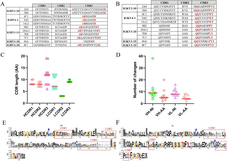
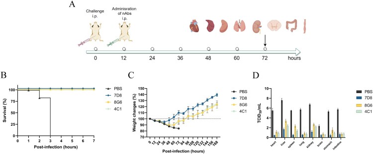
Ebola disease is a lethal viral hemorrhagic fever caused by ebolaviruses within the Filoviridae family with mortality rates of up to 90%. Monoclonal antibody (mAb) based therapies have shown great potential for the treatment of EVD. However, the potential emerging ebolavirus isolates and the negative effect of decoy protein on the therapeutic efficacy of antibodies highlight the necessity of developing novel antibodies to counter the threat of Ebola. Here, 11 fully human mAbs were isolated from transgenic mice immunized with GP protein and recombinant vesicular stomatitis virus-bearing GP (rVSV-EBOV GP). These mAbs were divided into five groups according to their germline genes and exhibited differential binding activities and neutralization capabilities. In particular, mAbs 8G6, 2A4, and 5H4 were cross-reactive and bound at least three ebolavirus glycoproteins. mAb 4C1 not only exhibited neutralizing activity but no cross-reaction with sGP. mAb 7D8 exhibited the strongest neutralizing capacity. Further analysis on the critical residues for the bindings of 4C1 and 8G6 to GPs was conducted using antibodies complementarity-determining regions (CDRs) alanine scanning. It has been shown that light chain CDR3 played a crucial role in binding and neutralization and that any mutation in CDRs could not improve the binding of 4C1 to sGP. Importantly, mAbs 7D8, 8G6, and 4C1 provided complete protections against EBOV infection in a hamster lethal challenge model when administered 12 h post-infection. These results support mAbs 7D8, 8G6, and 4C1 as potent antibody candidates for further investigations and pave the way for further developments of therapies and vaccines.

Keywords: Ebola virus; Ebola virus disease; fully human antibody; neutralizing antibodies; surrogate model; transgenic mice.


Conflict of interest statement

No potential conflict of interest was reported by the author(s).

Figures

Figure 1.
Figure 1.
Isolation of GP-specific antibodies from immunized transgenic mice. (A) Immunization strategy of antibody-humanized transgenic mice, CAMouse. Each mouse was immunized with five injections of antigens comprised of EBOV GPΔmuc and rVSV-EBOV GP at 2-week intervals starting on day 0. And 1 week after each immunization, blood was collected for the measurement of serum-neutralizing antibody titres. (B) The neutralizing antibody titres of the sera of GP-immunized CAMouse, 7093, 7094, 7095, 7103, and 7104, were assessed against rVSV-EBOV GP eGFP. (C) The binding of Abs in the supernatant’s individual in vitro expanded hybridoma cells (shown with dots) to EBOV GPΔTM was assessed by ELISA.
Figure 2.
Figure 2.
Analysis of EBOV GP-specific antibody sequences. (A, B) Antibodies with the same VH (A), and VL (B) germline genes are grouped and CDR1, CDR2, and CDR3 are listed. Bold and red letters indicate identical or conserved amino acids. (C) CDRs length of VH and VL genes. (D) Number of nucleotide (Nt) and amino acids (AA) mutations in VH and VL genes. (E, F) Multiple alignment of VH, and VL of mAbs depicted by WebLogo 3. CDR regions are marked.
Figure 3.
Figure 3.
Binding and neutralization profile of 11 fully human monoclonal antibodies. (A) Reactivity of mAbs to the indicated GPΔTM proteins, and truncated EBOV GPs determined by ELISA. (B) mAbs mediated neutralization of rVSV-EBOV GP eGFP. (C) Kinetics of 8G6, 4C1 9A9, 5A12, and 7D8 bindings analysed by BLI. On-rate (Kon), off-rate (Koff), and KD values for each GP ligand are shown below the sensograms.
Figure 4.
Figure 4.
Epitope analysis of 7D8, 8G6, and 4C1. (A) The crystal structure of EBOV GPΔmuc (PDB: 5JQ3) and structures of mAb variable regions were used to build the complex using the HDOCK SERVER software and analyse with Pymol. The chain A and B of EBOV GPΔmuc were coloured green and dark red, respectively. The VH and VL of nAbs were coloured light blue and blue, respectively. (B) OD450 values of the antibodies binding to truncated EBOV GPs. (C) The bindings of 4C1, 7D8, and 8G6 to truncated GPs were verified by Western Blot.
Figure 5.
Figure 5.
4C1 heavy- and light-chain gene sequence, critical residues for GPs recognition. (A) Alignment of 4C1 HC and LC and respective inferred human germline sequences. (B) Alanine scanning mutants of 4C1 HC and LC CDRs were assessed for binding affinity for EBOV GPΔTM and EBOV sGP relative to the WT 4C1. (C) Summary of 4C1 HC and LC CDR critical residues for bindings for EBOV GPΔTM and EBOV sGP. Mutated residues with values < 0.33, and > 3 were considered for GP binding. (D) Mutants were assessed for the neutralizing capacity for rVSV-EBOV GP eGFP relative to the WT 4C1.
Figure 6.
Figure 6.
8G6 heavy- and light-chain gene sequence, critical residues for GPs recognition. (A) Alignment of 8G6 HC and LC and respective inferred human germline sequences. (B) Alanine scanning mutants of 8G6 HC and LC CDRs were assessed for binding affinity for EBOV GPΔTM SUDV GPΔTM, BDBV GPΔTM, and TAFV GPΔTM relative to the WT 8G6. (C) Summary of 8G6 HC and LC CDR critical residues for bindings for EBOV GPΔTM, SUDV GPΔTM, BDBV GPΔTM, and TAFV GPΔTM. Mutated residues with values < 0.33, and > 3 were considered for GP binding. (D) Mutants were assessed for the neutralizing capacity for rVSV-EBOV GP eGFP relative to the WT 8G6.
Figure 7.
Figure 7.
Survival, weight change, and TCID50 of hamsters treated with mAbs. (A) Schematic representation of the therapeutic efficacy of antibodies evaluated in the hamster model (B) Hamsters (n = 6 per group) were inoculated with virus, and treated with 25 mg/kg of antibodies or PBS at 12 h post-infection, and monitored for 7 days. (C) Weight change of hamsters after virus infection and mAbs treatment. Hamsters were monitored for 7 days. (D) Virus titres (TCID50) of organs harvested at 3 days post infection.

Similar articles

References

    1. BRèS P. The epidemic of Ebola haemorrhagic fever in Sudan and Zaire, 1976: introductory note [J]. Bull World Health Organ. 1978;56(2):245. - PMC - PubMed
    1. Ebola haemorrhagic fever in Zaire, 1976 [J]. Bull World Health Organ. 1978;56(2):271–293. - PMC - PubMed
    1. Goldstein T, Anthony SJ, Gbakima A, et al. . The discovery of Bombali virus adds further support for bats as hosts of ebolaviruses [J]. Nat Microbiol. 2018;3(10):1084–1089. doi:10.1038/s41564-018-0227-2 - DOI - PMC - PubMed
    1. Ghosh S, Saha A, Samanta S, et al. . Genome structure and genetic diversity in the Ebola virus [J]. Curr Opin Pharmacol. 2021;60:83–90. doi:10.1016/j.coph.2021.06.010 - DOI - PubMed
    1. Jacob ST, Crozier I, Fischer WA, et al. . Ebola virus disease [J]. Nat Rev Dis Primers. 2020;6(1):13. doi:10.1038/s41572-020-0147-3 - DOI - PMC - PubMed