Case Reports
. 2024 Aug 9:2024:1595717.
doi: 10.1155/2024/1595717. eCollection 2024.

Two Pediatric Cases of Primary Ciliary Dyskinesia Caused by Loss-of-Function Variants in Oral-Facial-Digital Syndrome Gene, OFD1

Affiliations
Case Reports

Two Pediatric Cases of Primary Ciliary Dyskinesia Caused by Loss-of-Function Variants in Oral-Facial-Digital Syndrome Gene, OFD1

Yifei Xu et al. Case Rep Genet. .

Abstract

Primary ciliary dyskinesia (PCD) is a hereditary disease caused by genes related to motile cilia. We report two male pediatric cases of PCD caused by hemizygous pathogenic variants in the OFD1 centriole and centriolar satellite protein (OFD1) gene. The variants were NM_003611.3: c.[2789_2793delTAAAA] (p.[Ile930LysfsTer8]) in Case 1 and c.[2632_2635delGAAG] (p.[Glu878LysfsTer9]) in Case 2. Both cases had characteristic recurrent respiratory infections. Neither case had symptoms of oral-facial-digital syndrome type I. We identified a variant (c.2632_2635delGAAG) that has not been previously reported in any case of OFD1-PCD.


Conflict of interest statement

The authors declare that they have no conflicts of interest.

Figures

Figure 1
Figure 1
Imaging findings in Case 1. (a) Chest X-ray at 2 months of age showing situs solitus and consolidation (black arrow) with a loss of lung volume in the right upper lobe. (b) Chest computed tomography showing consolidation (black arrow) with an air bronchogram in the right upper lobe. (c) Transmission electron microscopy findings of a biopsy specimen from the nasal mucosa of case 1 showing normal ciliary axoneme ultrastructure. The cilium consists of one pair of central microtubules (white arrow) surrounded by nine pairs of well-arranged peripheral microtubules. The outer dynein arms (arrowheads) are presented.
Figure 2
Figure 2
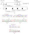
Family pedigree of case 1 with the results of Sanger sequencing. (a) Family pedigree of case 1. III-2 is the proband (Case 1) and his mother (II-3) is a carrier. III-3, -4, and -5 had symptoms similar to those of III-2, but genetic analysis has not been performed on these individuals. I-4 and II-5 are presumed to be carriers. (b) The proband (III-2) has a hemizygous variant in OFD1. (c) His father (II-2) does not carry the same variant. (d) The proband's mother (II-3) is a carrier of the same heterozygous deletion.
Figure 3
Figure 3
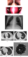
Imaging findings in Case 2. Ear endoscopy showing thickened bilateral tympanic membranes without a cone of light in the (a) right and (b) left ears. The middle nasal meatus is occluded with mucus secretion (white arrows) in the (c) right and (d) left nasal cavities. (e) Chest X-ray at 4 months of age showing situs inversus totalis and increased bronchovascular shadows (black arrow). Chest computed tomography of the (f) upper, (g) middle, and (h) lower lobes showing consolidation with ground glass opacity (black arrow) in the right lower lobe. (i) Transmission electron microscopy findings of Case 2. The cilium consists of one pair of central microtubules (white arrow) surrounded by nine pairs of well-arranged peripheral microtubules. The outer dynein arms (arrowheads) are presented.
Figure 4
Figure 4
Family pedigree of Case 2 with the results of Sanger sequencing. (a) Family pedigree of Case 2. III-2 is the proband. The parents are not carriers. (b) The proband (III-2) has a de novo deletion variant in OFD1. (c) Neither his father (II-1) nor (d) mother (II-2) carries the same variant.

References

    1. Takeuchi K., Kitano M., Ishinaga H., et al. Recent advances in primary ciliary dyskinesia. Auris Nasus Larynx . 2016;43(3):229–236. doi: 10.1016/j.anl.2015.09.012. - DOI - PubMed
    1. Zariwala M. A., Knowles M. R., Leigh M. W. Primary Ciliary Dyskinesia . Seattle, WA, USA: University of Washington Press; 2007. - PubMed
    1. Ferrante M. I., Feather S. A., Bulfone A., et al. Identification of the gene for oral-facial-digital type I syndrome. The American Journal of Human Genetics . 2001;68(3):569–576. doi: 10.1086/318802. - DOI - PMC - PubMed
    1. Macca M., Franco B. The molecular basis of oral-facial-digital syndrome, type 1. American Journal of Medical Genetics Part C: Seminars in Medical Genetics . 2009;(4):318–325. doi: 10.1002/ajmg.c.30224. - DOI - PubMed
    1. Alby C., Boutaud L., Bonnière M., et al. In utero ultrasound diagnosis of corpus callosum agenesis leading to the identification of orofaciodigital type 1 syndrome in female fetuses. Birth Defects Research . 2018;110(4):382–389. doi: 10.1002/bdr2.1154. - DOI - PubMed

Publication types

LinkOut - more resources