. 2024 May 16;9(8):2514-2526.
doi: 10.1016/j.ekir.2024.05.007. eCollection 2024 Aug.

Renal and Extrarenal Phenotypes in Patients With HNF1B Variants and Chromosome 17q12 Microdeletions

Collaborators, Affiliations

Renal and Extrarenal Phenotypes in Patients With HNF1B Variants and Chromosome 17q12 Microdeletions

Bénédicte Buffin-Meyer et al. Kidney Int Rep. .

Abstract

Introduction: Hepatocyte nuclear factor 1-beta (HNF1B) gene variants or the chromosome 17q12 deletion (17q12del) represent the most common monogenic cause of developmental kidney disease. Although neurodevelopmental disorders have been associated with the 17q12del, specific genotype-phenotype associations with respect to kidney function evolution have not yet been fully defined. Here, we aimed to determine whether 17q12del or specific HNF1B variants were associated with kidney survival in a large patient population with HNF1B disease.

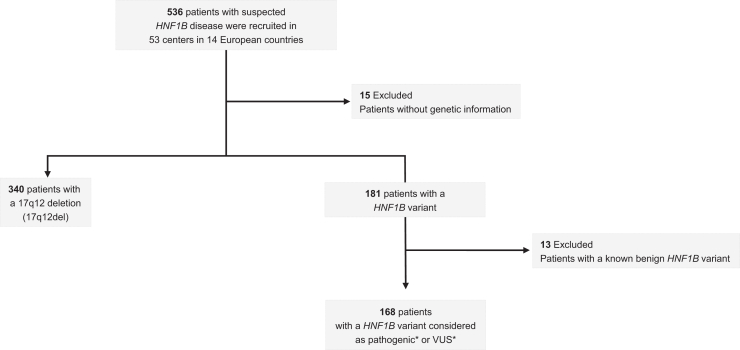
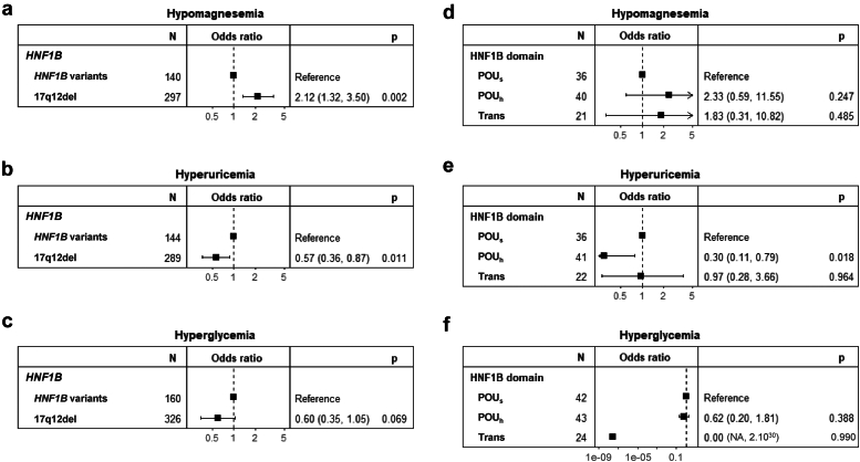
Methods: This was a retrospective observational study involving 521 patients with HNF1B disease from 14 countries using the European Reference Network for rare kidney diseases with detailed information on the HNF1B genotype (HNF1B variants or the 17q12del). Median follow-up time was 11 years with 6 visits per patient. The primary end point was progression to chronic kidney disease (CKD) stage 3 (estimated glomerular filtration rate [eGFR] < 60 ml/min per 1.73 m2). Secondary end points were the development of hypomagnesemia or extrarenal disorders, including hyperuricemia and hyperglycemia.

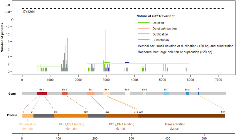
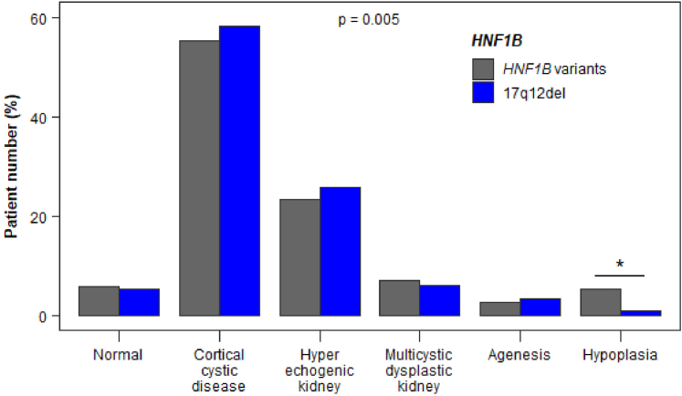
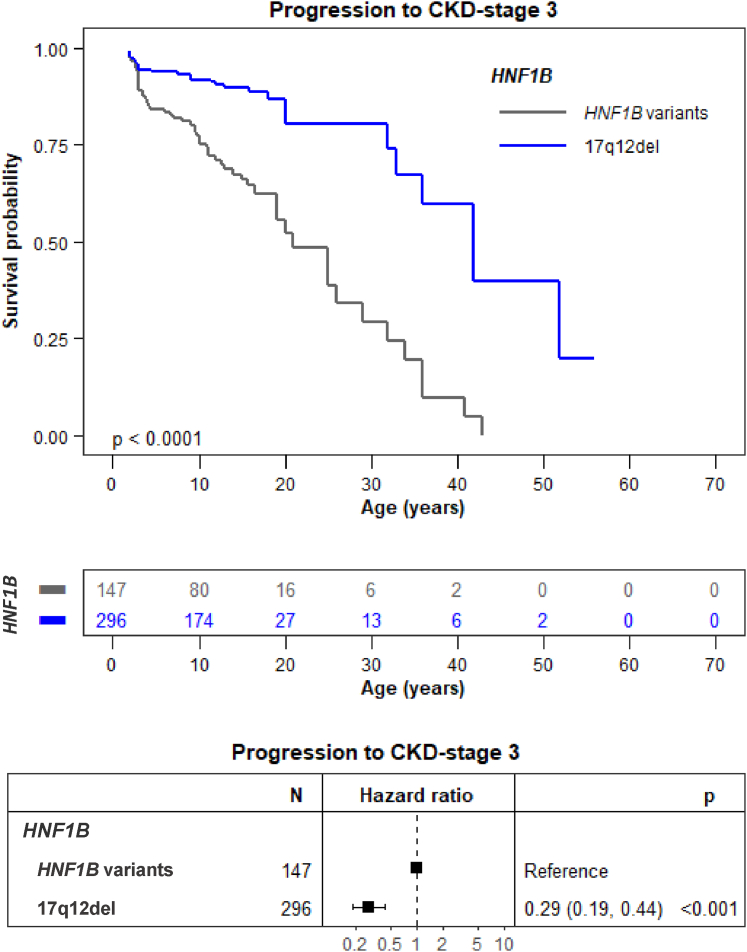
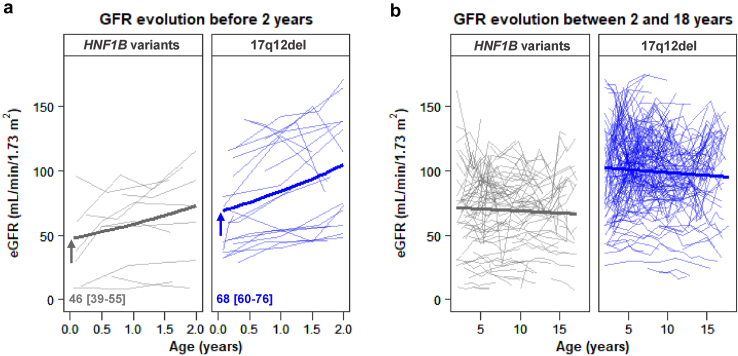
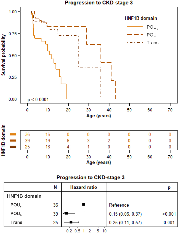
Results: Progression toward CKD stage 3 was significantly delayed in patients with the 17q12del compared to patients with HNF1B variants (hazard ratio [HR]: 0.29, 95% confidence interval [CI]: 0.19-0.44, P < 0.001). Progression toward CKD stage 3 was also significantly delayed when HNF1B variants involved the HNF1B Pit-1, Oct-1, and Unc-86 homeodomain (POUh) DNA-binding and transactivation domains rather than the POU-specific domain (POUs) DNA-binding domain (HR: 0.15 [95% CI: 0.06-0.37), P < 0.001 and HR: 0.25 (95% CI: 0.11-0.57), P = 0.001, respectively). Finally, the 17q12del was positively associated with hypomagnesemia and negatively associated with hyperuricemia, but not with hyperglycemia.

Conclusion: Patients with the 17q12del display a significantly better kidney survival than patients with other HNF1B variants; and for the latter, variants in the POUs DNA-binding domain lead to the poorest kidney survival. These are clinically relevant HNF1B kidney genotype-phenotype correlations that inform genetic counseling.

Keywords: HNF1B disease; chronic kidney disease; genotype-phenotype correlation; outcome.


Figures

None
Graphical abstract
Figure 1
Figure 1
Overview of patient recruitment and patient exclusion. ∗Definition of pathogenicity or variant of unknown significance (VUS) was based on the merger of the 3 following databases. ClinVar (https://www.ncbi.nlm.nih.gov/clinvar (db accessed July 11, 2023); LOVD (https://databases.lovd.nl/shared/variants/HNF1B#object_id=VariantOnTranscript%2CVariantOnGenome&id=HNF1B&order=VariantOnTranscript%2FDNA%2CASC&search_transcriptid=00009498&search_VariantOnTranscript/DNA=c.738G%3ET&page_size=100&page=1 (db accessed July 11, 2023)) and Leipzig_University (https://www.hnf1b.org (db accessed July 11, 2023). A variant was marked as pathogenic if in at least 1 database the variant was labelled “pathogenic”. In all other cases a variant was labelled “VUS.”
Figure 2
Figure 2
Position of molecular modifications on the HNF1B gene and HNF1B protein in 508 patients with HNF1B disease. Domains in the HNF1B protein were positioned according to.
Figure 3
Figure 3
Kidney ultrasound characteristics at diagnosis in the 521 patients with HNF1B variants. A global significant effect (P = 0.005) of the HNF1B genotype was observed. The adjusted standardized residual was equal to 4.80 for hypoplasia in patients with HNF1B variants, thereby indicating (≥3, see statistical analysis) that there were more patients with hypoplasia than would be expected by chance. In contrast, the adjusted standardized residual was equal to −6.99 for hypoplasia in patients with the 17q12del, thereby indicating (≤ −3, see statistical analysis) that there were less patients with hypoplasia than would be expected by chance. This strongly suggests that the risk of having hypoplasia was significantly lower in patients with the 17q12del than in patients with HNF1B variants (∗).
Figure 4
Figure 4
Progression to CKD stage 3 of patients the 17q12del compared to HNF1B variants. Progression to CKD is significantly delayed in patients with the 17q12del compared to patients with HNF1B variants (HR: 0.29 [95% CI: 0.19–0.44], P < 0.001). The survival curve was generated using data from children aged ≥ 2 years to dismiss changes in eGFR in early life. The point in time of progression to CKD stage 3 (eGFR < 60 ml/min per 1.73 m2) was entered as the chronological age of each patient. We considered that patients entered the study (baseline) at birth given the fact the HNF1B disease is a congenital nephropathy. The log-rank test for difference in survival yielded a P-value < 0.0001, indicating that the patients with 17q12del and HNF1B variants differed significantly in progression toward CKD stage 3. CI, confidence interval; CKD, chronic kidney disease; eGFR, estimated glomerular filtration rate; HR, hazard ratio.
Figure 5
Figure 5
Impact of the HNF1B genotype on eGFR trajectories in the pediatric period. Comparison of eGFR trajectories between the HNF1B variants and the 17q12del (a) before 2 years (10 and 21 patients for HNF1B variants and the 17q12del, respectively) and (b) between 2 and 18 years (91 and 175 patients for HNF1B variants and the 17q12del, respectively). Patients with <3 eGFR measurements during the period of interest were excluded. Individual and mean (bold) trajectories are plotted. The arrows and values indicate the mean (95% CI) eGFR at 1 week after birth. CI, confidence interval; eGFR, estimated glomerular filtration rate.
Figure 6
Figure 6
Impact of variants in the different HNF1B domains on the progression to CKD stage 3. Variants located in the POUh and transactivation domains displayed a significantly delayed progression toward CKD stage 3 compared to patients with variants in the POUs domain (HR: 0.15 [95% CI: 0.06–0.37], P < 0.001 and HR: 0.25 [95% CI: 0.11–0.57], P = 0.001, respectively). The survival curve was generated using data from children aged ≥2 years to dismiss changes in eGFR in early life. The point in time of progression to CKD stage 3 (eGFR < 60 ml/min per 1.73 m2) was entered as the chronological age of each patient. We considered that patients entered the study (baseline) at birth given that HNF1B disease is a congenital nephropathy. CI, confidence interval; CKD, chronic kidney disease; eGFR, estimated glomerular filtration rate; HR, hazard ratio; Trans, transactivation domain. The log rank test for difference in survival yielded a P-value < 0.0001, indicating that the patients with variants in the 3 HNF1B domains differed significantly in progression toward CKD stage 3.
Figure 7
Figure 7
The 17q12del and HNF1B variants in hypomagnesemia or extrarenal disorders. (a) Hypomagnesemia (magnesium < 0.6 mmol/l) was more frequent in patients with the 17q12del compared to patients with HNF1B variants. (b) Hyperuricemia (uric acid > 320 μmol/l) was less frequent in patients with the 17q12del compared to patients with HNF1B variants. (c) Hyperglycemia (fasting blood glucose > 1.26 g/l) was not different in the patient groups. (d) Hypomagnesemia was not different between patients with variants in the POUs, POUh, and transactivation domains. (e) Hyperuricemia was less frequent in patients with variants in the POUh than in the POUs and transactivation domains. (f) Hyperglycemia was not different between patients with variants in the POUs, POUh, and transactivation domains.

References

    1. Decramer S., Parant O., Beaufils S., et al. Anomalies of the TCF2 gene are the main cause of fetal bilateral hyperechogenic kidneys. J Am Soc Nephrol. 2007;18:923–933. doi: 10.1681/ASN.2006091057. - DOI - PubMed
    1. Weber S., Moriniere V., Knuppel T., et al. Prevalence of mutations in renal developmental genes in children with renal hypodysplasia: results of the Escape study. J Am Soc Nephrol. 2006;17:2864–2870. doi: 10.1681/ASN.2006030277. - DOI - PubMed
    1. Thomas R., Sanna-Cherchi S., Warady B.A., Furth S.L., Kaskel F.J., Gharavi A.G. HNF1B and PAX2 mutations are a common cause of renal hypodysplasia in the CKiD cohort. Pediatr Nephrol. 2011;26:897–903. doi: 10.1007/s00467-011-1826-9. - DOI - PMC - PubMed
    1. Heidet L., Decramer S., Pawtowski A., et al. Spectrum of HNF1B mutations in a large cohort of patients who harbor renal diseases. Clin J Am Soc Nephrol. 2010;5:1079–1090. doi: 10.2215/CJN.06810909. - DOI - PMC - PubMed
    1. Bellanné-Chantelot C., Chauveau D., Gautier J.F., et al. Clinical spectrum associated with hepatocyte nuclear factor-1beta mutations. Ann Intern Med. 2004;140:510–517. doi: 10.7326/0003-4819-140-7-200404060-00009. - DOI - PubMed

LinkOut - more resources