. 2024 May 15;9(8):2462-2473.
doi: 10.1016/j.ekir.2024.05.011. eCollection 2024 Aug.

Clinicopathologic Features of IgG4-Related Kidney Disease

Affiliations

Clinicopathologic Features of IgG4-Related Kidney Disease

Alessia Buglioni et al. Kidney Int Rep. .

Abstract

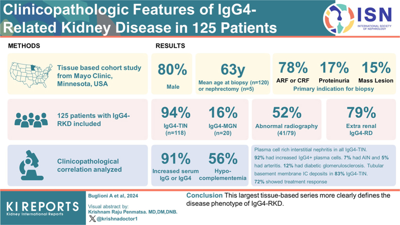
Introduction: IgG4-related disease (IgG4-RD) is a systemic immune-mediated disease that can involve nearly any organ. IgG4-RD can affect the kidney in different disease patterns, collectively referred to as IgG4-related kidney disease (IgG4-RKD).

Methods: We conducted a tissue-based cohort study with clinicopathological correlation in 125 patients with IgG4-RKD.

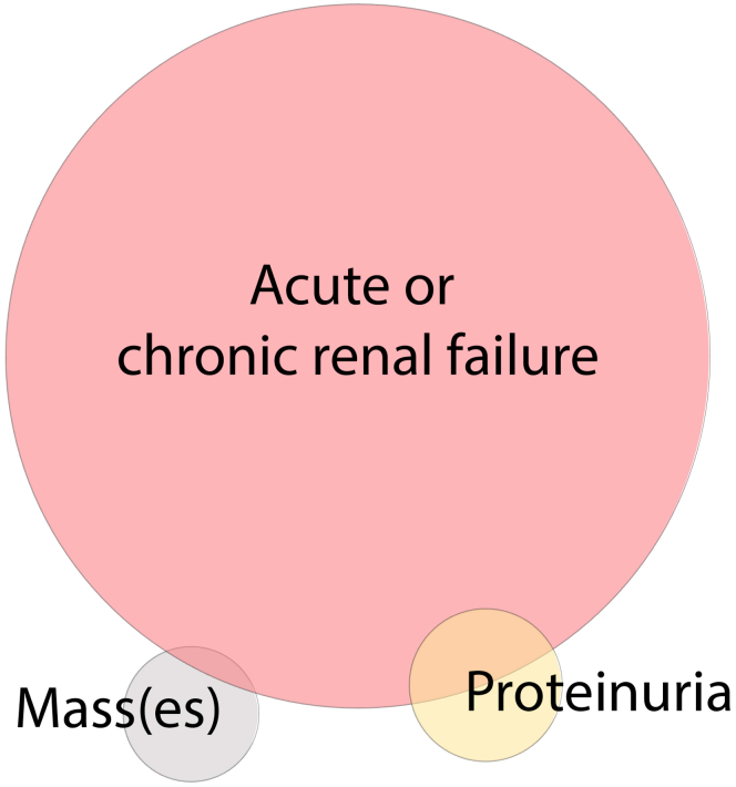
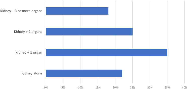
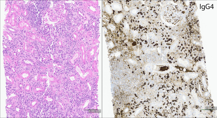
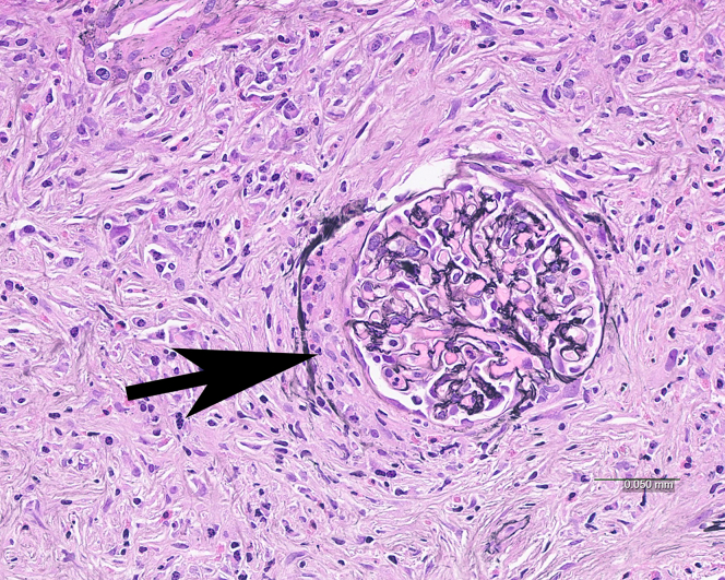
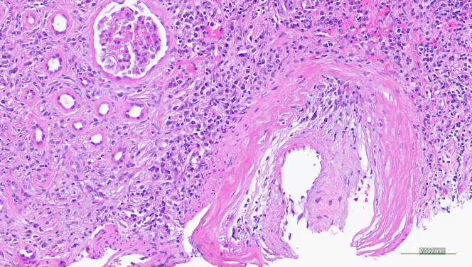
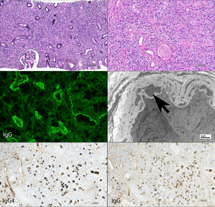
Results: The mean age at biopsy (n = 120) or nephrectomy (n = 5) was 63 years; 80% were male. One hundred eighteen patients (94%) had IgG4-related tubulointerstitial nephritis (IgG4-TIN); 20 patients (16%) had IgG4-related membranous glomerulonephritis (IgG4-MGN; 13 with concurrent IgG4-TIN). The primary clinical indication for biopsy/nephrectomy was acute or chronic renal failure in 78%, proteinuria in 17%, and mass lesion(s) in 15% (with overlap in primary indication). Fifty-two percent patients (41/79) had abnormal radiographic findings, including masses in 30% (24/79). All patients with IgG4-MGN had proteinuria. Extrarenal involvement by IgG4-RD was present in 79%. Median serum creatinine at presentation was 2.5 mg/dl (range 0.7-12). Serum IgG and/or IgG4 was increased in 91% (53/58); hypocomplementemia was present in 56% (43/77). Light microscopy showed plasma cell-rich interstitial nephritis in all cases of IgG4-TIN. Ninety-two percent of patients showed increased IgG4+ plasma cells. Seven percent showed an acute interstitial nephritis (AIN) pattern, and 5% showed non-necrotizing arteritis. Tubular basement membrane immune deposits were present in 83% of IgG4-TIN. Treatment information was available for 71 patients; 62 were treated with immunosuppression. Of those with elevated creatinine, 72% (41/57) showed a treatment response.

Conclusion: This largest tissue-based series more clearly defines the disease phenotype of IgG4-RKD.

Keywords: IgG4-related disease; autoimmune; interstitial nephritis; membranous nephropathy; tubulointerstitial nephritis.


Figures

None
Graphical abstract
Figure 1
Figure 1
Primary indication for biopsy/nephrectomy in IgG4-RKD. Distribution of primary indication for diagnostic procedure in our cohort. Most patients underwent biopsy for acute and/or chronic renal failure, while a smaller percentage underwent biopsy/nephrectomy for mass lesion(s) or proteinuria, with some overlap in primary indication. The size of the circles and degree of overlap is proportional to the number of patients in each category.
Figure 2
Figure 2
Multi-organ involvement in IgG4-RKD. Number of organs involved besides kidney in IgG4-RKD.
Figure 3
Figure 3
AIN pattern of IgG4-TIN. Plasma cell-rich interstitial inflammation with minimal fibrosis and no tubular atrophy (left panel). Immunoperoxidase staining for IgG4 (right panel) shows a marked increase in IgG4+ plasma cells. This pattern of injury shows minimal or no TBM immune deposits. (Hematoxylin and eosin).
Figure 4
Figure 4
Pseudocrescent in IgG4-TIN. Interstitial fibrosis and inflammation extend into and beyond the Bowman capsule of the glomerulus causing a crescent-like pattern of injury (arrow). No fibrinoid necrosis or glomerular basement membrane disruption is present. (Jones methenamine silver).
Figure 5
Figure 5
Plasma cell arteritis in IgG4-TIN. An artery shows numerous plasma cells intermixed with mononuclear cells within the thickened arterial intima. No fibrinoid necrosis is present. Note the concurrent plasma cell rich TIN pattern. Some cases of arteritis in IgG4-TIN showed mononuclear cells without significant plasma cells. (Hematoxylin and eosin).
Figure 6
Figure 6
Typical appearance of IgG4-TIN. A Jones-methenamine silver stain shows an expanded interstitium by a fibroinflammatory process (upper left panel). On higher magnification, the infiltrate is composed of numerous plasma cells, mononuclear cells, and several eosinophils (H & E, upper right panel). By immunofluorescence, there is granular tubular basement membrane and Bowman's capsule staining for IgG (middle left panel). By electron microscopy, amorphous electron dense deposits (arrow) are seen within the tubular basement membrane. The two bottom panels photograph of IgG4 and IgG immunoperoxidase stains of the same specimen in the same area on the slide: this example shows a marked increase in IgG4 positive plasma cells and also numerous IgG positive plasma cells, resulting in a high IgG4+ to IgG+ plasma cell ratio.

Similar articles

Cited by

References

    1. Wallace Z.S., Deshpande V., Mattoo H., et al. IgG4-related disease: clinical and laboratory features in one hundred twenty-five patients. Arthritis Rheumatol. 2015;67:2466–2475. doi: 10.1002/art.39205. - DOI - PMC - PubMed
    1. Wallace Z.S., Zhang Y., Perugino C.A., et al. Clinical phenotypes of IgG4-related disease: an analysis of two international cross-sectional cohorts. Ann Rheum Dis. 2019;78:406–412. doi: 10.1136/annrheumdis-2018-214603. - DOI - PMC - PubMed
    1. Umehara H., Okazaki K., Masaki Y., et al. A novel clinical entity, IgG4-related disease (IgG4RD): general concept and details. Mod Rheumatol. 2012;22:1–14. doi: 10.1007/s10165-011-0508-6. - DOI - PMC - PubMed
    1. Stone J.H., Zen Y., Deshpande V. IgG4-related disease. N Engl J Med. 2012;366:539–551. doi: 10.1056/NEJMra1104650. - DOI - PubMed
    1. Cornell L.D., Chicano S.L., Deshpande V., et al. Pseudotumors due to IgG4 immune-complex tubulointerstitial nephritis associated with autoimmune pancreatocentric disease. Am J Surg Pathol. 2007;31:1586–1597. doi: 10.1097/PAS.0b013e318059b87c. - DOI - PubMed

LinkOut - more resources