. 2024 Jul 9;27(8):110379.
doi: 10.1016/j.isci.2024.110379. eCollection 2024 Aug 16.

CCR3 knockdown attenuates prolonged underwater operations-induced cognitive impairment via alleviating microglia-mediated neuroinflammation

Affiliations

CCR3 knockdown attenuates prolonged underwater operations-induced cognitive impairment via alleviating microglia-mediated neuroinflammation

Houyu Zhao et al. iScience. .

Abstract

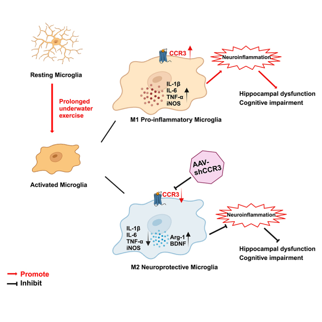
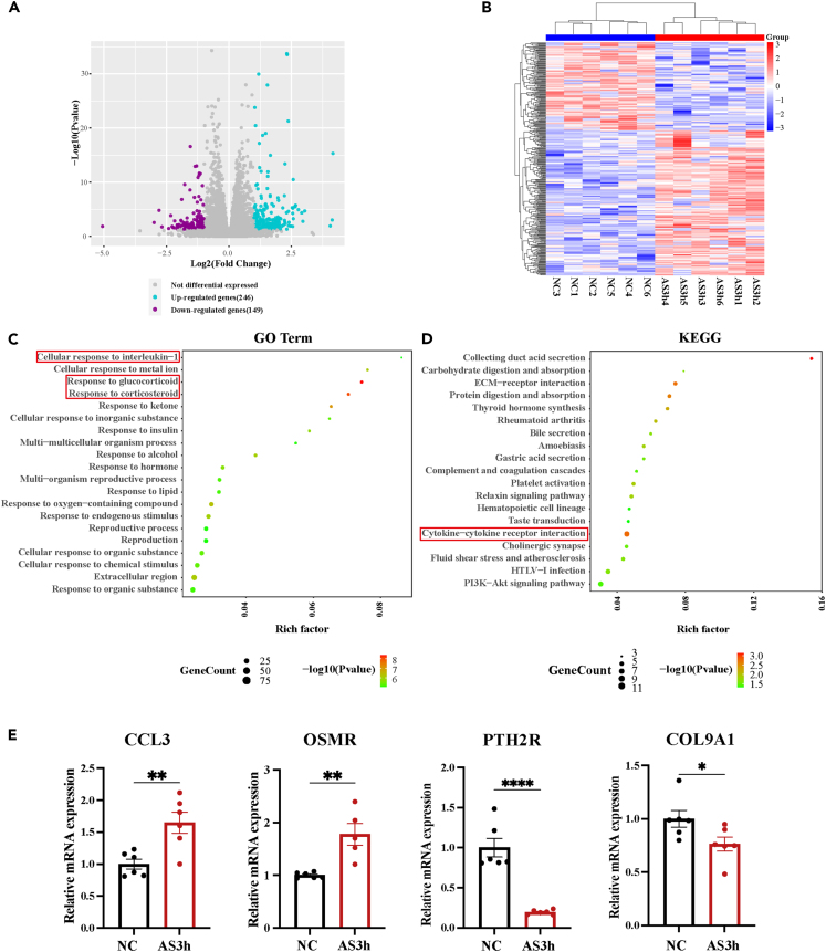
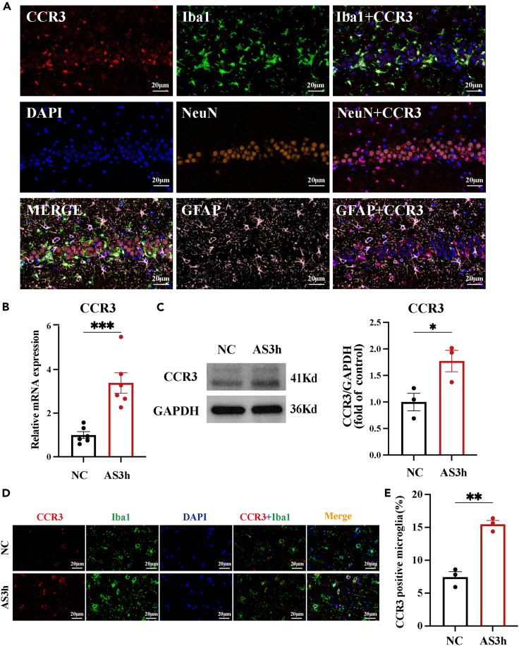
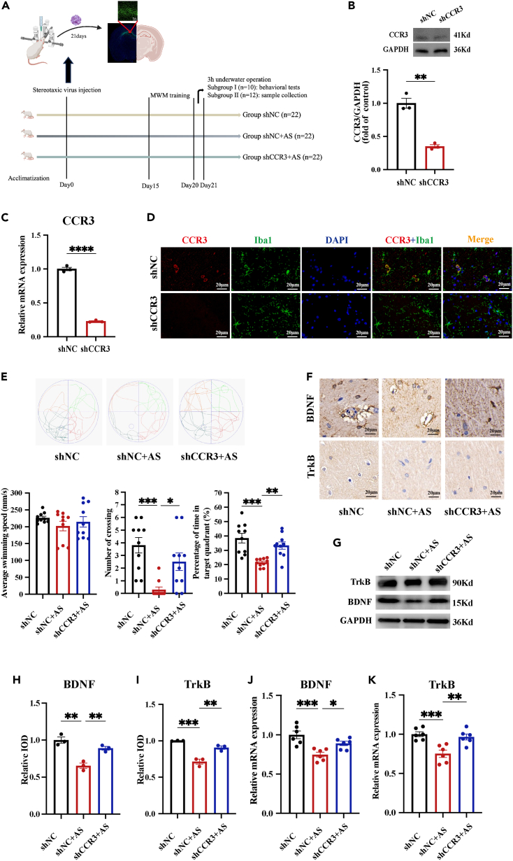
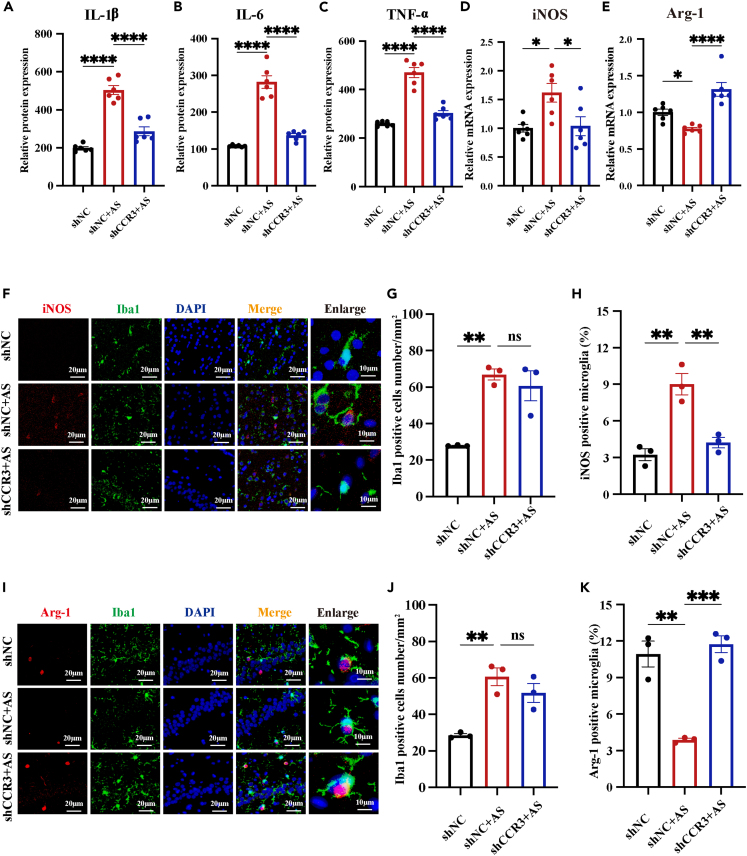
Maintaining cognitive integrity is crucial during underwater operations, which can significantly impact work performance and risk severe accidents. However, the cognitive effects of underwater operations and their underlying mechanism remain elusive, posing great challenges to the medical protection of professionals concerned. Here, we found that a single underwater operation session affects cognition in a time-dependent model. Prolonged exposure elicits significant cognitive impairment and hippocampal dysfunction, accompanied by increased neuroinflammation. Furthermore, RNA sequencing (RNA-seq) analysis revealed the involvement of neuroinflammation and highlighted the critical role of CCR3. Knockdown of CCR3 significantly rescued cognitive impairment and hippocampal dysfunction and reversed the upregulation of pro-inflammatory cytokines, by switching the activated microglia from a pro-inflammatory to a neuroprotective phenotype. Taken together, these results highlighted the time-dependent effects of a single underwater operation session on cognitive function. Knocking down CCR3 can attenuate neuroinflammation by regulating polarization of activated microglia, thereby alleviating prolonged underwater operations-induced cognitive impairment.

Keywords: Biological sciences; Neuroscience; Sensory neuroscience; Systems neuroscience.


Conflict of interest statement

The authors declare no competing interests.

Figures

None
Graphical abstract
Figure 1
Figure 1
Underwater operations affect cognitive function and prolonged exposure leads to cognitive decline (A) Schematic of the human experimental flowchart. Stress response, anxiety, and cognitive performance of special operation divers were evaluated before and after 3 single underwater operations of different durations (0.5 h, 1 h, 3 h respectively). (B) Concentration of cortisol in saliva before and after 3 single underwater operations. (C) Scores of anxiety before and after 3 single underwater operations. (D) Scores of memory before and after 3 single underwater operations. (E) Scores of processing speed before and after 3 single underwater operations. Data are expressed as mean ± SEM, statistical analysis is performed using paired two-tailed Student’s t test. ∗p < 0.05, ∗∗p < 0.01, ∗∗∗p < 0.001.
Figure 2
Figure 2
Prolonged underwater exercise elicits significant cognitive impairment in rats (A) Schematic of the animal experimental protocol. (B) The effect of underwater exercise on the concentration of CORT in serum of rats was detected using ELISA method. (n = 6 in each group). (C and E–G) The effect of underwater exercise on anxiety of rats was detected using OFT. (n = 10 in each group). Movement trajectory of rats in the OFT (C). Average moving speed of rats in the OFT (E). Total distance traveled by rats in the OFT (F). Time rats stayed in central areas in the OFT (G). (D and H–J) The effect of underwater exercise on cognitive performance of rats was detected using MWM. (n = 10 in each group). Movement trajectory of rats in the MWM (D). Average swimming speed of rats in the MWM (H). Numbers rats crossing the platform in the MWM (I). Percentage of time rats stayed in the target quadrant in the MWM (J). Data are expressed as mean ± SEM, statistical analysis is performed using one-way ANOVA followed by least significant difference (LSD) multiple comparisons test; compared with the group NC. ∗p < 0.05, ∗∗p < 0.01, ∗∗∗p < 0.001, ∗∗∗∗p < 0.0001.
Figure 3
Figure 3
Prolonged underwater exercise led to hippocampal dysfunction and neuroinflammation (A and B) The effect of underwater exercise on the morphology of hippocampus was detected using H&E and Nissl staining. (n = 3 in each group. Scale bar, 50μm). (C–E) The effect of underwater exercise on the protein expression of BDNF/TrkB in the hippocampus was detected using IHC staining. (n = 3 in each group. Scale bar, 20μm). (F and G) The effect of underwater exercise on the mRNA expression of BDNF/TrkB in the hippocampus was detected using RT-qPCR. (n = 6 in each group). (H) The effect of underwater exercise on the protein expression of BDNF/TrkB in the hippocampus was detected using western blotting (n = 3 in each group). (I and J) The effect of underwater exercise on the microglia activation was detected using IF staining. (n = 3 in each group. Scale bar, 20μm). (K–M) The effect of underwater exercise on the concentration of IL-1β, IL-6, and TNF-α in the hippocampus was detected using ELISA. (n = 6 in each group). Data are expressed as mean ± SEM, statistical analysis is performed using one-way ANOVE followed by LSD multiple comparisons test; compared with the group NC. ∗p < 0.05, ∗∗p < 0.01, ∗∗∗p < 0.001, ∗∗∗∗p < 0.0001.
Figure 4
Figure 4
RNA-seq identifies the involvement of neuroinflammation and the critical role of CCR3 (A) Clustered heatmap for DEGs from NC group and AS3h group (n = 6 in each group). (B) Volcano plot of differentially expressed genes in NC and AS3h group (purple, downregulation; blue, upregulation; gray, no change). (C) GO analysis of DEGs in NC and AS3h group. (D) KEGG analysis of DEGs in NC and AS3h group. (E) RT-qPCR of 4 selected DEGs to verify the results of RNA-seq. Data are expressed as mean ± SEM, statistical analysis is performed using unpaired two-tailed Student’s t test; compared with the group NC. ∗p < 0.05, ∗∗p < 0.01, ∗∗∗p < 0.001, ∗∗∗∗p < 0.0001.
Figure 5
Figure 5
CCR3 expression level was increased in the hippocampus after prolonged underwater exercise (A) Co-staining of CCR3 (red) and Iba1 (green), NeuN (orange), and GFAP (pink) in the hippocampus using IF staining (n = 3 in each group, Scale bar, 20μm). (B) The effect of prolonged underwater exercise on the mRNA expression of CCR3 in the hippocampus was detected using RT-qPCR (n = 6 in each group). (C) The effect of prolonged underwater exercise on the protein expression of CCR3 in the hippocampus was detected using western blotting (n = 3 in each group). (D and E) The effect of prolonged underwater exercise on the protein expression of CCR3 in microglia in the hippocampus was detected using IF staining (n = 3 in each group, Scale bar, 20μm). Data are expressed as mean ± SEM, statistical analysis is performed using unpaired two-tailed Student’s t test; compared with the group NC. ∗p < 0.05, ∗∗p < 0.01, ∗∗∗p < 0.001, ∗∗∗p < 0.0001.
Figure 6
Figure 6
CCR3 knockdown alleviates prolonged underwater exercise-induced cognitive impairment (A) Schematic of the knockdown experiment protocol. shNC and shCCR3 virus were injected into the hippocampus of rats and transfection efficiency was detected 3 weeks after injection. MWM training was conducted 15 days after virus injection and MWM test and sample collection were conducted after 3 h underwater exercise at the day 21 after virus injection. (B and C) The effect of AAV-CCR3 on the mRNA and protein levels of CCR3 in the hippocampus was detected by RT-qPCR (B) and western blot (C). (n = 3 in each group). (D) The effect of AAV-CCR3 on the protein levels of CCR3 in microglia in the hippocampus was detected by IF (Co-staining of CCR3 (red) and Iba1 (green)). (n = 3 in each group, Scale bar, 20μm). (E) The effect of AAV-CCR3 on cognitive performance of rats was detected by the MWM test. Movement trajectories of rats in the MWM. (n = 10 in each group). (F, H, and I) The effect of AAV-CCR3 on the protein expression of BDNF/TrkB of rats was detected by IHC. (n = 3 in each group, Scale bar, 20μm). (G) The effect of underwater exercise on the protein expression of BDNF/TrkB in the hippocampus was detected using western blotting (n = 3 in each group). (J and K). The effect of AAV-CCR3 on the mRNA expression of BDNF/TrkB of rats was detected by RT-qPCR. (n = 6 in each group). Data are expressed as mean ± SEM, statistical analysis is performed using unpaired two-tailed Student’s t test (B-C), or one-way ANOVA followed by Tukey’s multiple comparisons test (F–K); p < 0.05, p < 0.01, p < 0.001, p < 0.0001; “#” compared with the group shNC, “∗” compared with the group shNC+AS.
Figure 7
Figure 7
CCR3 knockdown alleviated neuroinflammation induced by prolonged underwater exercise through regulating microglia polarization (A–C) The effect of AAV-CCR3 on the Concentration of IL-1β, IL-6, and TNF-α in the hippocampus was detected by ELISA (n = 6 in each group). (D and E) The effect of AAV-CCR3 on the mRNA levels of Arg-1 and iNOS in the hippocampus was detected by RT-qPCR (n = 6 in each group). (F–K) The effect of AAV-CCR3 on the protein levels of Arg-1 and iNOS in microglia in the hippocampus was detected by IF staining (n = 3 in each group, Scale bar, 20μm). Co-staining of iNOS (red) and Iba1 (green) in the hippocampus (F). Iba1 positive cells number in the hippocampus (G). Percentage of iNOS positive microglia in the hippocampus (H). Co-staining of Arg-1 (red) and Iba1 (green) in the hippocampus (I). Iba1 positive cells number in the hippocampus (J). Percentage of Arg-1positive microglia in the hippocampus (G). Data are expressed as mean ± SEM, statistical analysis is performed using one-way ANOVA followed by Tukey’s multiple comparisons test; p < 0.05, p < 0.01, p < 0.001, p < 0.0001; # compared with the group shNC, ∗ compared with the group shNC+AS.

Similar articles

References

    1. de Jong F.J.M., Wingelaar T.T., van Hulst R.A. Pulmonary oxygen toxicity in occupational diving. Occup. Med. 2023;73:231–232. doi: 10.1093/occmed/kqad043. - DOI - PMC - PubMed
    1. Kelly K.R., Palombo L.J., Jensen A.E., Bernards J.R. Efficacy of closed cell wet-suit at various depths and gas mixtures for thermoprotection during military training dives. Front. Physiol. 2023;14 doi: 10.3389/fphys.2023.1165196. - DOI - PMC - PubMed
    1. Diamond A. Executive functions. Annu. Rev. Psychol. 2013;64:135–168. doi: 10.1146/annurev-psych-113011-143750. - DOI - PMC - PubMed
    1. Steinberg F., Doppelmayr M. Executive Functions of Divers Are Selectively Impaired at 20-Meter Water Depth. Front. Psychol. 2017;8:1000. doi: 10.3389/fpsyg.2017.01000. - DOI - PMC - PubMed
    1. Hammerton Z. Determining the variables that influence SCUBA diving impacts in eastern Australian marine parks. Ocean Coast Manag. 2017;142:209–217.