

Changes of the peripapillary vascular parameters in premature infants without retinopathy of prematurity using U-net segmentation
- PMID: 39156772
- PMCID: PMC11286446
- DOI: 10.18240/ijo.2024.08.10
Changes of the peripapillary vascular parameters in premature infants without retinopathy of prematurity using U-net segmentation
Abstract
Aim: To quantitatively assess the changes in mean vascular tortuosity (mVT) and mean vascular width (mVW) around the optic disc and their correlation with gestational age (GA) and birth weight (BW) in premature infants without retinopathy of prematurity (ROP).
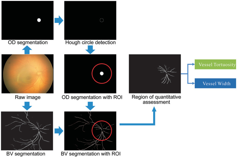
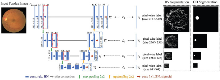
Methods: A single-center retrospective study included a total of 133 (133 eyes) premature infants [mean corrected gestational age (CGA) 43.6wk] without ROP as the premature group and 130 (130 eyes) CGA-matched full-term infants as the control group. The peripapillary mVT and mVW were quantitatively measured using computer-assisted techniques.
Results: Premature infants had significantly higher mVT (P=0.0032) and lower mVW (P=0.0086) by 2.68 (104 cm-3) and 1.85 µm, respectively. Subgroup analysis with GA showed significant differences (P=0.0244) in mVT between the early preterm and middle to late preterm groups, but the differences between mVW were not significant (P=0.6652). The results of the multiple linear regression model showed a significant negative correlation between GA and BW with mVT after adjusting sex and CGA (P=0.0211 and P=0.0006, respectively). For each day increase in GA at birth, mVT decreased by 0.1281 (104 cm-3) and for each 1 g increase in BW, mVT decreased by 0.006 (104 cm-3). However, GA (P=0.9402) and BW (P=0.7275) were not significantly correlated with mVW.
Conclusion: Preterm birth significantly affects the peripapillary vascular parameters that indicate higher mVT and narrower mVW in premature infants without ROP. Alterations in these parameters may provide new insights into the pathogenesis of ocular vascular disease.
Keywords: computer-assisted techniques; premature infants; retinal vessels parameter; retinopathy of prematurity.
International Journal of Ophthalmology Press.
Conflict of interest statement
Conflicts of Interest: Liu S, None; Liu L, None; Ma CX, None; Huang LH, None; Li B, None.
Figures
Similar articles
-
Association between retinal vascular fractal dimensions and retinopathy of prematurity: an AI-assisted retrospective case-control study.Int Ophthalmol. 2025 Mar 18;45(1):105. doi: 10.1007/s10792-025-03461-1. Int Ophthalmol. 2025. PMID: 40100468
-
Incidence and risk factors for retinopathy of prematurity in premature, extremely low birth weight and extremely low gestational age infants.BMC Ophthalmol. 2022 Sep 13;22(1):367. doi: 10.1186/s12886-022-02591-9. BMC Ophthalmol. 2022. PMID: 36096834 Free PMC article.
-
Risk Factors Associated with Retinopathy of Prematurity in Very and Extremely Preterm Infants.Medicina (Kaunas). 2021 Apr 27;57(5):420. doi: 10.3390/medicina57050420. Medicina (Kaunas). 2021. PMID: 33925286 Free PMC article.
-
[Characteristics and associated factors of early refractive parameters in premature infants].Zhonghua Yan Ke Za Zhi. 2021 May 11;57(5):353-357. doi: 10.3760/cma.j.cn112142-20200427-00288. Zhonghua Yan Ke Za Zhi. 2021. PMID: 33915638 Chinese.
-
Peripapillary Choroidal Thickness in Former Preterm and Full-Term Infants Aged From 4 to 10 Years.Invest Ophthalmol Vis Sci. 2016 Dec 1;57(15):6548-6553. doi: 10.1167/iovs.16-20128. Invest Ophthalmol Vis Sci. 2016. PMID: 27918828
References
-
- Gupta K, Campbell JP, Taylor S, Brown JM, Ostmo S, Paul Chan RVP, Dy J, Erdogmus D, Ioannidis S, Kalpathy-Cramer J, Kim SJ, Chiang MF, Imaging and Informatics in Retinopathy of Prematurity Consortium A quantitative severity scale for retinopathy of prematurity using deep learning to monitor disease regression after treatment. JAMA Ophthalmol. 2019;137(9):1029–1036. - PMC - PubMed
-
- Taylor S, Brown JM, Gupta K, Campbell JP, Ostmo S, Paul Chan RVP, Dy J, Erdogmus D, Ioannidis S, Kim SJ, Kalpathy-Cramer J, Chiang MF, Imaging and Informatics in Retinopathy of Prematurity Consortium Monitoring disease progression with a quantitative severity scale for retinopathy of prematurity using deep learning. JAMA Ophthalmol. 2019;137(9):1022–1028. - PMC - PubMed
LinkOut - more resources
Full Text Sources
Research Materials