

A surgical alternative of fusiform penetrating keratoplasty for the management of severe infectious keratitis
- PMID: 39156785
- PMCID: PMC11286445
- DOI: 10.18240/ijo.2024.08.07
A surgical alternative of fusiform penetrating keratoplasty for the management of severe infectious keratitis
Abstract
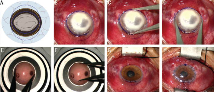
Aim: To describe the surgical procedure of fusiform penetrating keratoplasty (FPK) using multiple trephines of different sizes for treating patients with severe infectious keratitis.
Methods: Fourteen eyes underwent FPK, and 15 eyes received conventional penetrating keratoplasty (PK) were included in the study. The best-corrected visual acuity (BCVA), refractive outcomes, endothelial cell density, and postoperative complications were recorded.
Results: The FPK group was followed for an average of 15.3±2.1mo, whereas the PK group was followed for 16.1±1.9mo. The corneal ulcers were elliptical-shaped in all 14 eyes in the FPK group. The mean BCVA (logMAR, 0.26±0.13) showed no statistically significant differences from that in the PK group (logMAR, 0.21±0.12, P>0.05) at 1y after surgery. But the mean curvature, mean astigmatism, and mean spherical equivalent in the FPK group were lower than those in the PK group (P<0.05). Peripheral anterior synechia was observed in one patient in the FPK group, whereas 6 patients in the PK group. Suture loosening and neovascularization were observed in 4 and 5 eyes in the PK group, respectively. No graft immune rejection or elevation of intraocular pressure was observed in the two groups.
Conclusion: For patients with elliptical-shaped corneas or corneal ulcers, FPK can avoid disrupting of corneal limbus, reduce the risk of postoperative complications, and can result in satisfactory visual quality.
Keywords: cornea; fusiform penetrating keratoplasty; infectious keratitis; multiple trephines.
International Journal of Ophthalmology Press.
Conflict of interest statement
Conflicts of Interest: Qi XL, None; Wang LC, None; Wang ML, None; Gao H, None.
Figures


References
-
- Galvis V, Tello A, Laiton AN, Salcedo SLL. Indications and techniques of corneal transplantation in a referral center in Colombia, South America (2012–2016) Int Ophthalmol. 2019;39(8):1723–1733. - PubMed
-
- Pluzsik MT, Seitz B, Flockerzi FA, Langenbucher A, Tóth G, Bohle RM, Szentmáry N. Changing trends in penetrating keratoplasty indications between 2011 and 2018—histopathology of 2123 corneal buttons in a single center in Germany. Curr Eye Res. 2020;45(10):1199–1204. - PubMed
-
- Szentmáry N, Langenbucher A, Kus MM, Naumann GOH, Seitz B. Long-term refractive results of elliptical excimer laser penetrating keratoplasty (EELPK) Curr Eye Res. 2007;32(11):953–959. - PubMed
-
- Lang GK, Naumann GO, Koch JW. A new elliptical excision for corneal transplantation using an excimer laser. Arch Ophthalmol. 1990;108(7):914–915. - PubMed
LinkOut - more resources
Full Text Sources