

Testicular tumor in a case of, undescended testes, persistent mullerian duct syndrome and transverse testicular ectopia: Report of a case and review of the literature
- PMID: 39157013
- PMCID: PMC11327389
- DOI: 10.1016/j.eucr.2024.102803
Testicular tumor in a case of, undescended testes, persistent mullerian duct syndrome and transverse testicular ectopia: Report of a case and review of the literature
Abstract
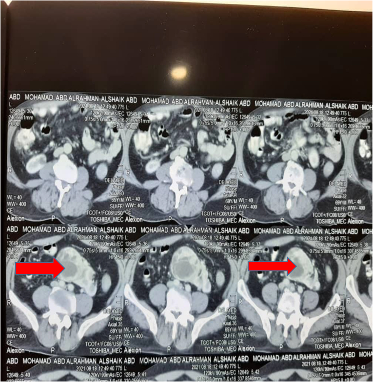
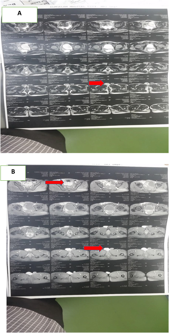
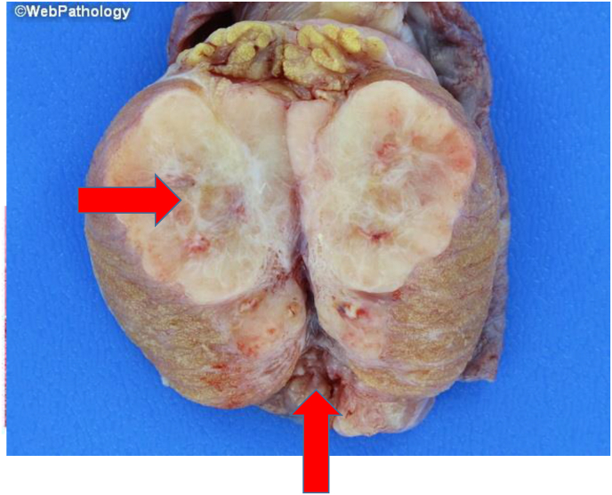
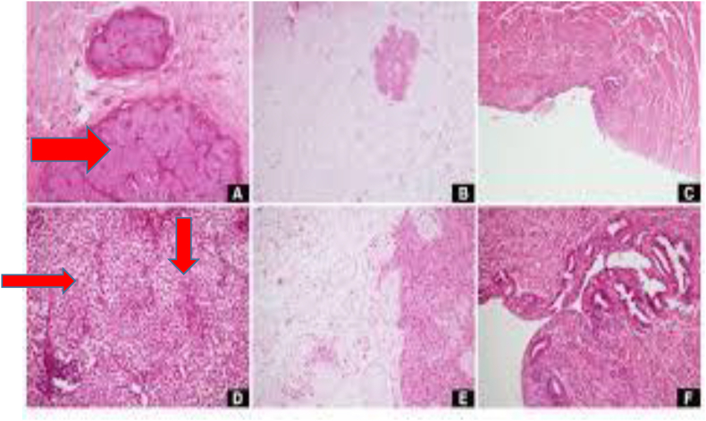
A 20-year-old with normal male body features and secondary sexual characteristics presented with a right testicular swelling. Imaging revealed a right testicular mass, leading to a diagnosis of classical seminoma. During inguinal orchiectomy, a solid testicular mass was found on the right side along with two spermatic cords, one attached to the mass and the other to a structure resembling a testes. Examination showed the presence of other testes and a rudimentary uterus, indicating a rare case of a testicular tumor coexisting with undescended testes and transverse testicular ectopia (TTE) in a Pseudohermaphrodite with "persistent mullerian duct syndrome" (PMDS).
Keywords: Disorder of sexual differentiation; Persistent mullerian duct; Testicular tumor; Transverse testicular ectopia.
© 2024 The Author.
Conflict of interest statement
I hereby declare that I have no conflict of interest with the journal, publisher, or reviewer. This statement is directed to the chief editor in charge.
Figures
References
-
- Yuksel B., Saygun O., Hengirmen S. Persistent müllerian duct syndrome associated with irreducible inguinal hernia, bilateral cryptorchidism and testicular neoplasia: a case report. Acta Chir Belg. 2006;106(1):119–120. - PubMed
-
- Prakash N., Khurana A., Narula B. Persistent Müllerian duct syndrome. Indian J Pathol Microbiol. 2009;52(4):546. - PubMed
-
- Nilson O. Hernia uteri inguinalis bein Manne. Acta Chir Scand. 1939;83:231–249.
-
- Von Lenhossek M. Ectopia testes transverse. Anat Anzeiger. 1886;1:376.
Publication types
LinkOut - more resources
Full Text Sources
