. 2024 Aug 2:12:1444846.
doi: 10.3389/fbioe.2024.1444846. eCollection 2024.

Based on mutated aptamer-smartphone colorimetric detection of metronidazole in milk

Affiliations

Based on mutated aptamer-smartphone colorimetric detection of metronidazole in milk

Sicheng Zhang et al. Front Bioeng Biotechnol. .

Abstract

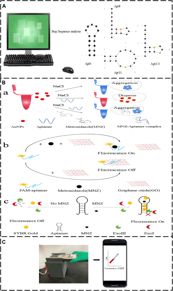
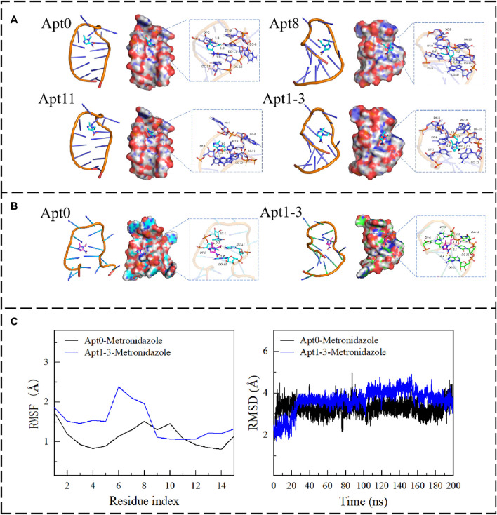
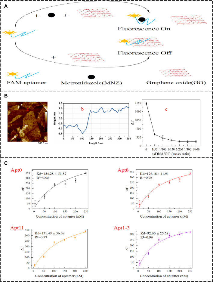
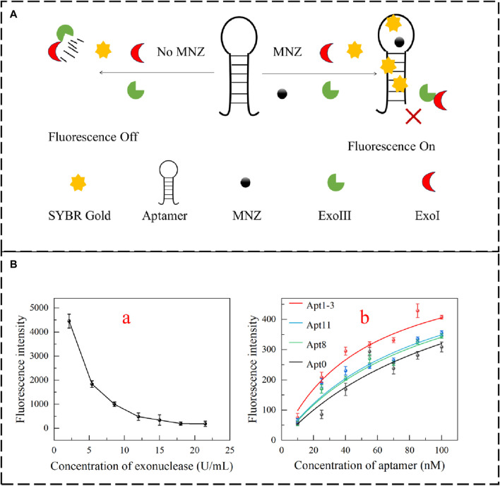
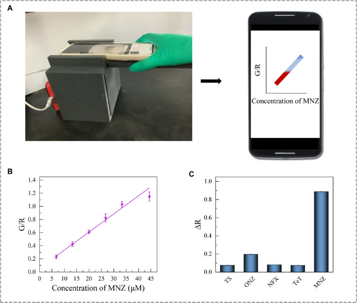
Excessive residue of metronidazole (MNZ) in food is harmful to the human body. There is an urgent demand to develop a portable tool for MNZ detection on-site. In this study, fifteen aptamers were prepared through targeted base mutation. Apt1-3 with the highest enrichment was chosen for further study. Its affinity was characterized by molecular docking simulation, AuNPs colorimetric assay, graphene oxide (GO) fluorescence assay, and exonuclease assay. Kd was determined by GO fluorescence assay (Kd: 92.60 ± 25.59 nM). Its specificity was also characterized by an exonuclease assay. A novel aptasensor was constructed by using the newly identified aptamer combined with the smartphone dark box. The principle of color change is caused by the aggregation state of AuNPs. Smartphones act as reading instruments. The detection can be completed in just a few seconds without the aid of instruments, achieving a detection limit of 0.15 nmol/mL and a range of 6.7-44.4 nmol/mL (R 2 = 0.9810). Therefore, the constructed smartphone colorimetric sensor based on mutant aptamers has important applications in food detection.

Keywords: aptamer; base mutation; colorimetric; metronidazole; smartphone.


Conflict of interest statement

The authors declare that the research was conducted in the absence of any commercial or financial relationships that could be construed as a potential conflict of interest.

Figures

FIGURE 1
FIGURE 1
(A) The original aptamer-Apt0 was mutated to Apt8, Apt11 and Apt1-3. (B,a) Aptamer AuNPs schematic. (B,b) Graphene oxide fluorescence method schematic diagram. (B,c) Exonuclease schematic diagram. (C) Smartphone Dark box colorimetric device.
FIGURE 2
FIGURE 2
(A) The binding mode of Apt0, Apt8, Apt11, and Apt1-3 with MNZ. (B) The binding mode of Apt0 and Apt1-3 with MNZ after MD. (C) The RMSF and RMSD of Apt0 and Apt3 after 200 ns of molecular dynamics.
FIGURE 3
FIGURE 3
(A) AuNPs colorimetric schematic. (B) Transmission electron microscopy (TEM) of AuNPs in different states of 100 nm. (C) Spectrograms of AuNPs in different states (a: newly prepared AuNPs; b: 0.06 μM aptamer and 40 mM NaCl were added; c: added 0.06 μM aptamer, 40 μM MNZ, and 40 mM NaCl; d: added 40 mM NaCl). (D) AuNPs size distribution map.
FIGURE 4
FIGURE 4
(A) GO-based fluorescence method to detect the affinity of aptamer schematic diagram. (B,a,b) The AFM images of GO. (B,c) Effect of GO addition concentration on △F. (C) Apt0, Apt8, Apt11, and Apt1-3 Kd value fitting curve.
FIGURE 5
FIGURE 5
(A) Schematic diagram for detecting the affinity of aptamers based on the exonuclease fluorescent method. (B,a) Optimization of exonuclease incorporation. (B,b) Apt0, Apt8, Apt11 and Apt1-3 fluorescence intensity fitting.
FIGURE 6
FIGURE 6
(A) Smartphone colorimetric device diagram and chart. (B) Apt1-3 Smartphone colorimetric linear range. (C) Apt1-3 proprietary validation chart.

Similar articles

References

    1. Alkhamis O., Canoura J., Ly P. T., Xiao Y. (2023). Using exonucleases for aptamer characterization, engineering, and sensing. Acc. Chem. Res. 56 (13), 1731–1743. 10.1021/acs.accounts.3c00113 - DOI - PubMed
    1. Bhalla N., Jolly P., Formisano N., Estrela P. (2016). Introduction to biosensors. Essays Biochem. 60 (1), 1–8. 10.1042/EBC20150001 - DOI - PMC - PubMed
    1. Canoura J., Alkhamis O., Liu Y., Willis C., Xiao Y. (2023). High-throughput quantitative binding analysis of DNA aptamers using exonucleases. Nucleic Acids Res. 51 (4), e19. 10.1093/nar/gkac1210 - DOI - PMC - PubMed
    1. Chen L., Yang G., Qu F. (2024). Advances of aptamer-based small-molecules sensors in body fluids detection. Talanta 268, 125348. 10.1016/j.talanta.2023.125348 - DOI - PubMed
    1. Chen X., Huang Y., Duan N., Wu S., Xia Y., Ma X., et al. (2014). Screening and identification of DNA aptamers against T-2 toxin assisted by graphene oxide. J. Agric. Food Chem. 62 (42), 10368–10374. 10.1021/jf5032058 - DOI - PubMed

LinkOut - more resources