. 2024 Jul 18:22:101792.
doi: 10.1016/j.bonr.2024.101792. eCollection 2024 Sep.

Vasorin-deficient mice display disturbed vitamin D and mineral homeostasis in combination with a low bone mass phenotype

Affiliations

Vasorin-deficient mice display disturbed vitamin D and mineral homeostasis in combination with a low bone mass phenotype

Marco Eijken et al. Bone Rep. .

Abstract

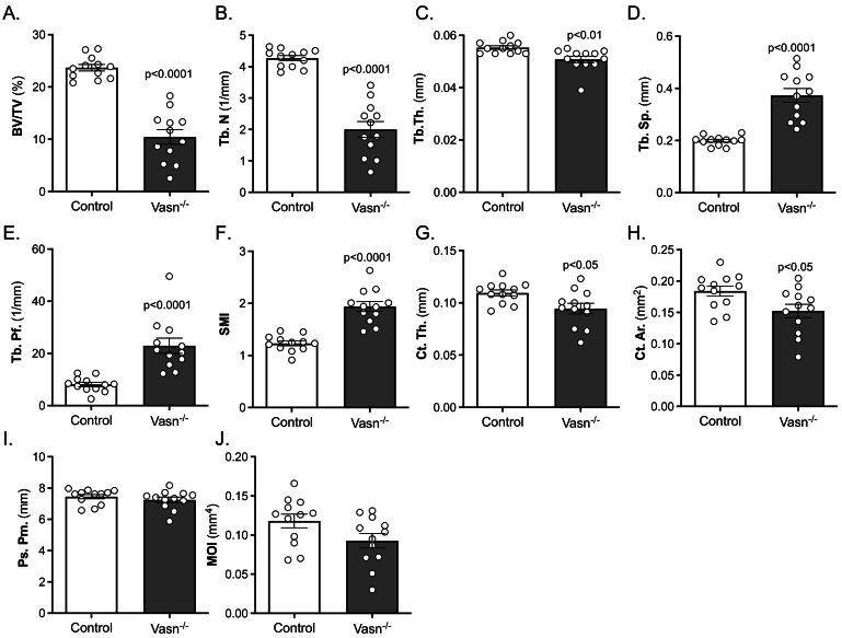
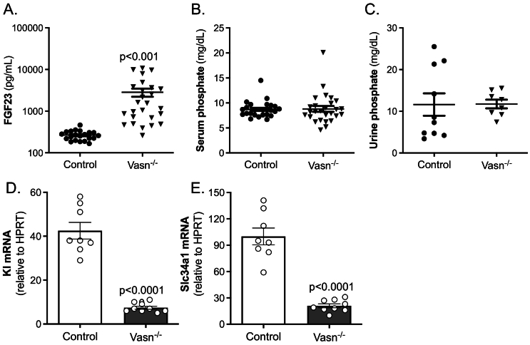
Vasorin (Vasn) is a pleiotropic molecule involved in various physiological and pathological conditions, including cancer. Vasn has also been detected in bone cells of developing skeletal tissues but no function for Vasn in bone metabolism has been implicated yet. Therefore, this study aimed to investigate if Vasn plays a significant role in bone biology. First, we investigated tissue distribution of Vasn expression, using lacZ knock-in reporter mice. We detected clear Vasn expression in skeletal elements of postnatal mice. In particular, osteocytes and bone forming osteoblasts showed high expression of Vasn, while the bone marrow was devoid of signal. Vasn knockout mice (Vasn -/- ) displayed postnatal growth retardation and died after four weeks. MicroCT analysis of femurs from 22- to 25-day-old Vasn -/- mice demonstrated reduced trabecular and cortical bone volume corresponding to a low bone mass phenotype. Ex vivo bone marrow cultures demonstrated that osteoclast differentiation and activity were not affected by Vasn deficiency. However, osteogenesis of Vasn -/- bone marrow cultures was disturbed, resulting in lower numbers of alkaline phosphate positive colonies, impaired mineralization and lower expression of osteoblast marker genes. In addition to the bone phenotype, these mice developed a vitamin D3-related phenotype with a strongly reduced circulating 25-hydroxyvitamin D3 and 1,25-dihydroxyvitamin D3 and urinary loss of vitamin D binding protein. In conclusion, Vasn-deficient mice suffer from severe disturbances in bone metabolism and mineral homeostasis.

Keywords: Bone; Osteoblast; Vasorin; Vitamin D; Vitamin D binding protein.


Conflict of interest statement

The authors declare that they have no known competing financial interests or personal relationships that could have appeared to influence the work reported in this paper.

Figures

Fig. 1
Fig. 1
LacZ reporter activity in a VasnlacZ embryonic day E15.5 embryo, indicating widespread expression of Vasn, predominantly in the skeleton (A). LacZ expression in periosteal osteoblasts and osteocytes (B) as well as trabecular osteoblasts (C) from 10-week-old VasnlacZ mice. Insets in B and C represent stainings on wild type mice. Vasn mRNA expression panel (D) for 2 samples of femoral bone marrow (slot 1 + 2), tibial bone marrow (slot 3 + 4), mouse bone (slot 5 + 6), mouse osteoclasts (slot 7 + 8), mouse osteoblasts (slot 9 + 10).
Fig. 2
Fig. 2
μCT analysis of wild type (Control) and Vasn−/− mice (n = 12) femurs demonstrating trabecular bone volume fraction (BV/TV) (A), trabecular number n(Tb.N) (B), trabecular thickness (Tb.Th) (C), trabecular spacing (Tb.Sp) (D), trabecular pattern factor (Tb.Pf) (E), structure model index (SMI) (F), cortical thickness (Ct.Th) (G), cortical area (Ct.Ar) (H), periosteal perimeter (Ps.Pm) (I) and moment of inertia (MOI) (J).
Fig. 3
Fig. 3
Bone formation (A) and resorption (B) markers total procollagen 1 N-terminal pro peptide (P1NP) and tartrate-resistant acid phosphatase (TRAP), respectively, determined in serum of wild type (control) and Vasn−/− mice (P1NP: n = 9–11; TRAP: n = 11–13).
Fig. 4
Fig. 4
Osteoclastogenesis and osteoblastogenesis of bone marrow cultures derived from control and Vasn−/− mice (n = 8). After 6 days of differentiation TRAP positive mono- bi- and multi-nucleated cells were counted (A) and bone resorption pits were stained and quantified (B). Osteoblast cultures were stained after 4, 6 and 9 days for quantification of alkaline phosphatase (ALP) positive colonies (C) and after 11, 14 and 17 days for mineralized colonies, using alizarin Red (D). *p < 0.05, **p < 0.01, ***p < 0.001.
Fig. 5
Fig. 5
Gene expression in bone marrow-derived osteoblast cultures from control and Vasn−/− mice. Osteoblastic mRNA expression of (A) Vasn, (B) Runx2, (C) Bglap (D) Col1a1, (E) Alpl, (F) Spp1, (G) Smad7, (H) Klf10, (I) Sod1, (J) Hif1a and (K) Vegf, all corrected by normalization for the housekeeping gene Hprt1 (n = 8–9).
Fig. 6
Fig. 6
Calcium homeostasis markers determined in serum of control and Vasn−/− mice. (A) 1,25(OH)2D3 (n = 6–8), (B) 25(OH)D3 (n = 6–8), (C) parathyroid hormone (PTH) (n = 23–26), (D) serum calcium, (E) urinary calcium (n = 14–17).
Fig. 7
Fig. 7
Vitamin D binding protein (DBP) measurements in serum and urine of control and Vasn−/− mice. (A) serum DBP, (B) urinary DBP (n = 10–13). Renal mRNA expression of (C) megalin (Lrp2) and (D) cubulin (Cubn), corrected by normalization for the housekeeping gene Hprt (n = 8–9).
Fig. 8
Fig. 8
Phosphate homeostasis markers in serum and urine of control and Vasn−/− mice. (A) serum Fgf23 (n = 26–29), (B) urinary phosphate (n = 8–18), (C) serum phosphate (n = 27–36). Renal mRNA expression of (C) klotho (Kl) and (D) sodium-dependent phosphate transport protein 2 A (Slc34a1), corrected by normalization for the housekeeping gene Hprt (n = 8–9).

References

    1. Andrique C., Bonnet A.L., Dang J., Lesieur J., Krautzberger A.M., Baroukh B., et al. Vasorin as an actor of bone turnover? J. Cell. Physiol. 2024 Mar 19. (Epub 20240319) - PubMed
    1. Bonnet A.L., Chaussain C., Broutin I., Rochefort G.Y., Schrewe H., Gaucher C. From vascular smooth muscle cells to Folliculogenesis: what about Vasorin? Front Med (Lausanne). 2018;5:335. (Epub 20181204) - PMC - PubMed
    1. Bouxsein M.L., Boyd S.K., Christiansen B.A., Guldberg R.E., Jepsen K.J., Muller R. Guidelines for assessment of bone microstructure in rodents using micro-computed tomography. J. Bone Miner. Res. 2010;25(7):1468–1486. Jul. (Epub 2010/06/10) - PubMed
    1. Dallas S.L., Prideaux M., Bonewald L.F. The osteocyte: an endocrine cell ... And more. Endocr. Rev. 2013;34(5):658–690. Oct. (Epub 20130423) - PMC - PubMed
    1. Dardenne O., Prud'homme J., Arabian A., Glorieux F.H., St-Arnaud R. Targeted inactivation of the 25-hydroxyvitamin D(3)-1(alpha)-hydroxylase gene (CYP27B1) creates an animal model of pseudovitamin D-deficiency rickets. Endocrinology. 2001;142(7):3135–3141. Jul. - PubMed

LinkOut - more resources