. 2024 Oct 24;144(17):1821-1833.
doi: 10.1182/blood.2023022257.

Coagulation factor XI regulates endothelial cell permeability and barrier function in vitro and in vivo

Affiliations

Coagulation factor XI regulates endothelial cell permeability and barrier function in vitro and in vivo

Cristina Puy et al. Blood. .

Abstract

Loss of endothelial barrier function contributes to the pathophysiology of many inflammatory diseases. Coagulation factor XI (FXI) plays a regulatory role in inflammation. Although activation of FXI increases vascular permeability in vivo, the mechanism by which FXI or its activated form FXIa disrupts endothelial barrier function is unknown. We investigated the role of FXIa in human umbilical vein endothelial cell (HUVEC) or human aortic endothelial cell (HAEC) permeability. The expression patterns of vascular endothelial (VE)-cadherin and other proteins of interest were examined by western blot or immunofluorescence. Endothelial cell permeability was analyzed by Transwell assay. We demonstrate that FXIa increases endothelial cell permeability by inducing cleavage of the VE-cadherin extracellular domain, releasing a soluble fragment. The activation of a disintegrin and metalloproteinase 10 (ADAM10) mediates the FXIa-dependent cleavage of VE-cadherin, because adding an ADAM10 inhibitor prevented the cleavage of VE-cadherin induced by FXIa. The binding of FXIa with plasminogen activator inhibitor 1 and very low-density lipoprotein receptor on HUVEC or HAEC surfaces activates vascular endothelial growth receptor factor 2 (VEGFR2). The activation of VEGFR2 triggers the mitogen-activated protein kinase (MAPK) signaling pathway and promotes the expression of active ADAM10 on the cell surface. In a pilot experiment using an established baboon model of sepsis, the inhibition of FXI activation significantly decreased the levels of soluble VE-cadherin to preserve barrier function. This study reveals a novel pathway by which FXIa regulates vascular permeability. The effect of FXIa on barrier function may be another way by which FXIa contributes to the development of inflammatory diseases.


Conflict of interest statement

Conflict-of-interest disclosure: C.U.L. and E.I.T. are employees of Aronora Inc, a company that may have a commercial interest in the results of this research. J.J.S. serves as a medical consultant for Aronora, Inc; this potential conflict of interest has been reviewed and managed by the Oregon Health and Science University conflict of interest in research committee. The remaining authors declare no competing financial interests.

Figures

None
Graphical abstract
Figure 1.
Figure 1.
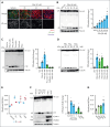
FXIa induces VE-cadherin shedding and EC permeability. (A) HUVECs were grown on gelatin-coated glass coverslips and incubated with FXIa (30 nM) for 2 to 6 hours. Cells were fixed and stained for VE-cadherin (green), actin (red), and nuclei (blue). Bar, 50 μm. (B) HUVECs were incubated with FXIa for 1 to 6 hours. Cells were lysed and analyzed by western blotting using an anti–C-terminal VE-cadherin antibody. Results are representative of 3 experiments. Significance (∗P < .05) determined by Kruskal-Wallis testing with Dunns correction for multiple comparisons. Data are mean ± standard error of the mean (SEM; n = 3). (C) HUVECs were incubated with FXIa (5 or 30 nM), thrombin (10 nM), kallikrein (30 nM), or FXIIa (30 nM) for 6 hours. Cells were lysed and analyzed by western blotting using an anti–C-terminal anti–VE-cadherin antibody. Results are representative of 3 experiments. Significance (∗P < .05) determined by Kruskal-Wallis testing with Dunns correction for multiple comparisons. Data are mean ± SEM (n = 3). (D) HUVECs were incubated with FXIa (30 nM), or FXa (10 or 30 nM), or FIXa (10 or 30 nM) for 6 hours. Cells were lysed and analyzed by western blotting using an anti–C-terminal VE-cadherin antibody. Results are representative of 3 experiments. Significance (∗P < .05) determined by Kruskal-Wallis testing with Dunns correction for multiple comparisons. Data are mean ± SEM (n = 3). (E) HUVECs were grown to confluence on gelatin-coated Transwell filters and incubated with thrombin, FXa, or FXIa (5 nM), for 2 or 6 hours. Permeability of Evans Blue-BSA was measured after 60 minutes of incubation. Data are mean ± SEM (n = 3). Significance (∗P < .05) was determined by Kruskal-Wallis testing with Dunns correction for multiple comparisons. (F) HUVECs were incubated with FXIa (1, 5, or 30 nM), TNFα (10 ng/mL), or VEGF (100 ng/mL) for 6 hours. Cells were lysed and analyzed by western blotting using an anti–C-terminal VE-cadherin, anti–ICAM-1, or anti–VCAM-1 antibodies. Results are representative of 3 experiments. Data are mean ± SEM. Significance (∗P < .05) was determined by Kruskal-Wallis testing with Dunns correction for multiple comparisons. (G) HUVECs were grown to confluence on gelatin-coated Transwell filters and incubated with FXIa (30 nM), VEGF (100 ng/mL), or TNFα (10 ng/mL) for 6 hours. Permeability of Evans Blue-BSA was measured after 60 minutes of incubation. Data are mean ± SEM (n = 3). Significance (∗P < .05) was determined by Kruskal-Wallis testing with Dunns correction for multiple comparisons.
Figure 2.
Figure 2.
Effect of ADAM10 inhibitor on VE-cadherin cleavage induced by FXIa. HUVECs were incubated with FXIa for 6 hours in the presence or absence of the serine protease inhibitor, PPACK (100 μM); the thrombin inhibitor, hirudin (25 μg/mL); or the ADAM10 inhibitor, GI254023X (10 μM). (A) Cells were lysed and analyzed by western blotting using an anti–C-terminal VE-cadherin antibody. Results are representative of 3 experiments. Significance (∗P < .05) determined by Kruskal-Wallis testing with Dunns correction for multiple comparisons. Data are mean ± SEM (n = 3). (B) Cell media were analyzed by using a sVE cadherin enzyme-linked immunosorbent assay. Data are mean ± SEM (n = 3). Significance (∗P < .05) determined by Kruskal-Wallis testing with Dunns correction for multiple comparisons. (C) Cell media were also analyzed by western blot using an anti–N-terminal VE-cadherin antibody. Results representative of 3 experiments. (D) HUVECs were grown on gelatin-coated glass coverslips and incubated with FXIa (30 nM) for 6 hours in the absence or presence of GI254023X (10 μM). Cells were fixed and stained for VE-cadherin (green), actin (red), and nuclei (blue). Bar, 50 μm. (E) HUVECs were grown to confluence on gelatin-coated Transwell filters and incubated with FXIa (30 nM) in the absence or presence of GI254023X (10 μM). Permeability of Evans Blue-BSA was measured after 60 minutes of incubation. Data are mean ± SEM (n = 4). Significance (∗P < .05) determined by Kruskal-Wallis testing with Dunns correction for multiple comparisons. (F) HUVECs were incubated with FXIa (30 nM) for 6 hours in the absence or presence of PPACK (100 μM). Cell surface was biotinylated and cell lysates were precipitated with NeutrAvidin agarose beads and probed with an anti-ADAM10, anti–N-terminal VE-cadherin, or anti-PECAM1 antibodies. Significance (∗P < .05) determined by Kruskal-Wallis testing with Dunns correction for multiple comparisons. Data are mean ± SEM (n = 3).
Figure 3.
Figure 3.
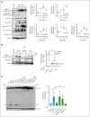
Role of the PI3K-Akt-eNOS and PLCγ1-ERK signaling pathways on VE-cadherin cleavage induced by FXIa. (A) HUVECs were incubated with FXIa (30 nM). Cells were lysed and immunoblotted with antibodies for phosphorylated ERK1/2 T202/Y204, p38 MAPK T180/Y182. Akt S473, eNOS S1177, PLCγ1 Y783, Src Y416, FAK Y397, or tubulin (n = 3). Significance (∗P < .05) determined by Kruskal-Wallis testing with Dunns correction for multiple comparisons. Data are mean ± SEM (n = 3). (B) HUVECs were incubated with FXIa, FXIa-PPACK, or FXI (30 nM) for 15 or 30 minutes. Cells were lysed and immunoblotted with antibodies for phosphorylated ERK1/2 T202/Y204 or Akt S473. Significance (∗P < .05) determined by Kruskal-Wallis testing with Dunns correction for multiple comparisons. Results are representative of 3 experiments. (C) HUVECs were incubated with vehicle (dimethyl sulfoxide [DMSO]) or FXIa for 6 hours in the absence or presence of the Src inhibitor, PP2 (10 μM); the protein kinase A (PKA) inhibitor, H-89 (10 μM); the eNOS inhibitor, L-NAME (5 μM); or the ERK1/2 inhibitor, LY3214996 (1 μM). Cells were lysed and analyzed by western blotting using an anti–C-terminal VE-cadherin antibody. Results are representative of 3 experiments. Significance (∗P < .05) determined by Kruskal-Wallis testing with Dunns correction for multiple comparisons. Data are mean ± SEM (n = 3).
Figure 4.
Figure 4.
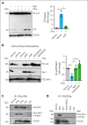
FXIa–PAI-1 complex interaction with VLDLR induces VE-cadherin shedding. (A) HUVECs were incubated with FXIa (30 nM) for 6 hours in the absence or presence of the low-density lipoprotein receptor antagonist, RAP (50 ng/mL). Cells were lysed and analyzed by western blotting using an anti–C-terminal VE-cadherin antibody. Significance (∗P < .05) determined by Kruskal-Wallis testing with Dunns correction for multiple comparisons. Data are mean ± SEM (n = 3). (B) HUVECs were incubated with FXIa (30 nM) in the absence or presence of PPACK (100 μM), GI254023X (5 μM), or RAP (50 ng/mL) for 6 hours. HUVECs cell surfaces were biotinylated, and cell lysates were precipitated with NeutrAvidin agarose beads. The precipitates were probed with an anti–N-terminal VE-cadherin or anti–PAI-1 antibodies. Significance (∗P < .05) determined by Kruskal-Wallis testing with Dunns correction for multiple comparisons. Data are mean ± SEM (n = 3). (C) HUVECs were incubated with FXIa (30 nM) at 4°C for 2 hours. Cells were washed and incubated with PPACK (100 μM) for 30 minutes; lysed in the presence of PPACK; followed by immunoprecipitation with an anti-FXI LC and western blotting with an anti–PAI-1, anti-VLDLR, or anti–platelet EC adhesion molecule 1 (PECAM-1) antibodies. Results are representative of 3 experiments. (D) HUVECs were incubated with FXIa, FXI, or FXIa-PPACK (30 nM) at 4°C for 2 hours in the absence or presence of RAP (50 ng/mL). Cells were washed and incubated with PPACK (100 μM) for 30 minutes, lysed in the presence of PPACK followed by with an anti-FXI light chain immunoprecipitation and western blotting with an anti–PAI-1 antibody or anti-VLDLR antibodies. Results are representative of 3 experiments.
Figure 5.
Figure 5.
Role of FXIa on Dab1 and VEGFR2 activation. (A) HUVECs were incubated with FXIa (30 nM) in the absence or presence of (A) the Src inhibitor, PP2 (10 μM), or the low-density lipoprotein receptor antagonist, RAP (50 ng/mL). Cells were lysed and immunoblotted with antibodies for Dab1 Y2020, PLCγ1 Y783, or tubulin. Results are representative of 3 experiments. Significance (∗P < .05) determined by Kruskal-Wallis testing with Dunns correction for multiple comparisons. (B) HUVECs were incubated with FXIa (30 nM) in the absence or presence of the VEGFR2 inhibitors, BFH772 (1 μM) or SU 1498 (5 μM). Cells were lysed and immunoblotted with antibodies for Dab1 Y2020, Src Y416, PLCγ1 Y783, or tubulin, or lysates were separated by Phos-tag sodium dodecyl sulfate–polyacrylamide gel electrophoresis and immunoblotted for VEGFR2 phosphorylation (n = 3). Significance (∗P < .05) determined by Kruskal-Wallis testing with Dunns correction for multiple comparisons. Data are mean ± SEM (n = 3). (C) HUVECs were incubated with vehicle or FXIa for 6 hours in the absence or presence of the VEGFR2 kinase activity inhibitor, SU1498 (5 μM), or the blocking anti-VEGF antibody, ramucirumab (10 μg/mL). Cells were lysed and analyzed by western blotting using an anti–C-terminal VE-cadherin antibody. Results are representative of 3 experiments. Significance (∗P < .05) determined by Kruskal-Wallis testing with Dunns correction for multiple comparisons. Data are mean ± SEM (n = 3). (D) HUVECs were grown to confluence on gelatin-coated Transwell filters and incubated with FXIa (30 nM), for 6 hours in the absence or presence of PP2 (10 μM) or SU1498 (5 μM). Permeability of Evans Blue-BSA was measured after 60 minutes of incubation. Data are mean ± SEM (n = 4). Significance (∗P < .05) determined by Kruskal-Wallis testing with Dunns correction for multiple comparisons. (E) HUVECs were incubated with FXIa (30 nM) for 6 hours in the absence or presence of the ADAM10 inhibitor, GI254023X (5 μM), or the Src inhibitor, PP2. Cell media were collected and immunoblotted with antibodies for N-terminal VE-cadherin or N-terminal VEGFR2. Results are representative of 3 experiments.
Figure 6.
Figure 6.
Effect of FXIa on EC permeability on HAECs. (A) HAECs were incubated with vehicle or FXIa for 6 hours in the absence or presence of the VEGFR2 kinase activity inhibitor, SU1498 (5 μM), the ADAM10 inhibitor, GI254023X (10 μM), or RAP (50 ng/mL). Cells were lysed and analyzed by western blotting using an anti–C-terminal VE-cadherin antibody. Results are representative of 3 experiments. Significance (∗P < .05) determined by Kruskal-Wallis testing with Dunns correction for multiple comparisons. (B) HAECs were grown to confluence on gelatin-coated Transwell filters and incubated with FXIa (30 nM) for 6 hours in the absence or presence of PP2 (10 μM), SU1498 (5 μM), or GI254023X (10 μM). Permeability of Evans Blue-BSA was measured after 60 minutes of incubation. Data are mean ± SEM (n = 4). Significance (∗P < .05) determined by Kruskal-Wallis testing with Dunns correction for multiple comparisons. (C) HAECs were incubated with FXIa (30 nM) for 15 to 60 minutes. Cells were lysed and immunoblotted with antibodies for phosphorylated Dab1 Y2020, Src Y416, VEGFR2 Y1175, ERK1/2 T202/Y204, eNOS S1177, PLCγ1 Y783, or tubulin. Results are representative of 3 experiments. Significance (∗P < .05) determined by Kruskal-Wallis testing with Dunns correction for multiple comparisons. Data are mean ± SEM (n = 3). (D) HAECs were incubated with FXIa (30 nM) for 15 to 60 minutes in the presence or absence of PP2 or SU1498. Cells were lysed and immunoblotted with antibodies for phosphorylated Dab1 Y2020, PLCγ1 Y783, or tubulin. Results are representative of 3 experiments. Significance (∗P < .05) determined by Kruskal-Wallis testing with Dunns correction for multiple comparisons. Data are mean ± SEM (n = 3).
Figure 7.
Figure 7.
Effect of the blocking anti-FXI antibody, 3G3, on sVE-cadherin levels after infusion of heat-inactivated S aureus into baboons. Time course change of sVE-cadherin levels. Data are mean ± standard deviation (SD) (n = 3). Same time points are compared between baboons challenged with 3 × 1010 colony forming units of S aureus per kg, and S aureus plus the blocking anti-FXI antibody, 3G3 (SA + 3G3) using 2-tailed student t test; ∗P < .05.

References

    1. Claesson-Welsh L, Dejana E, McDonald DM. Permeability of the endothelial barrier: identifying and reconciling controversies. Trends Mol Med. 2021;27(4):314–331. - PMC - PubMed
    1. Wettschureck N, Strilic B, Offermanns S. Passing the vascular barrier: endothelial signaling processes controlling extravasation. Physiol Rev. 2019;99(3):1467–1525. - PubMed
    1. Tzima E, Irani-Tehrani M, Kiosses WB, et al. A mechanosensory complex that mediates the endothelial cell response to fluid shear stress. Nature. 2005;437(7057):426–431. - PubMed
    1. Nawroth R, Poell G, Ranft A, et al. VE-PTP and VE-cadherin ectodomains interact to facilitate regulation of phosphorylation and cell contacts. EMBO J. 2002;21(18):4885–4895. - PMC - PubMed
    1. Lampugnani MG, Orsenigo F, Gagliani MC, Tacchetti C, Dejana E. Vascular endothelial cadherin controls VEGFR-2 internalization and signaling from intracellular compartments. J Cell Biol. 2006;174(4):593–604. - PMC - PubMed

Publication types

MeSH terms

LinkOut - more resources