. 2024 Aug 20;13(16):e035617.
doi: 10.1161/JAHA.124.035617. Epub 2024 Aug 19.

Ultrafast Cooling With Total Liquid Ventilation Mitigates Early Inflammatory Response and Offers Neuroprotection in a Porcine Model of Cardiac Arrest

Affiliations

Ultrafast Cooling With Total Liquid Ventilation Mitigates Early Inflammatory Response and Offers Neuroprotection in a Porcine Model of Cardiac Arrest

Yara Abi Zeid Daou et al. J Am Heart Assoc. .

Abstract

Background: Brain injury is one of the most serious complications after cardiac arrest (CA). To prevent this phenomenon, rapid cooling with total liquid ventilation (TLV) has been proposed in small animal models of CA (rabbits and piglets). Here, we aimed to determine whether hypothermic TLV can also offer neuroprotection and mitigate cerebral inflammatory response in large animals.

Methods and results: Anesthetized pigs were subjected to 14 minutes of ventricular fibrillation followed by cardiopulmonary resuscitation. After return of spontaneous circulation, animals were randomly subjected to normothermia (control group, n=8) or ultrafast cooling with TLV (TLV group, n=8). In the latter group, TLV was initiated within a window of 15 minutes after return of spontaneous circulation and allowed to reduce tympanic, esophageal, and bladder temperature to the 32 to 34 °C range within 30 minutes. After 45 minutes of TLV, gas ventilation was resumed, and hypothermia was maintained externally until 3 hours after CA, before rewarming using heat pads (0.5 °C-1 °C/h). After an additional period of progressive rewarming for 3 hours, animals were euthanized for brain withdrawal and histological analysis. At the end of the follow-up (ie, 6 hours after CA), histology showed reduced brain injury as witnessed by the reduced number of Fluroro-Jade C-positive cerebral degenerating neurons in TLV versus control. IL (interleukin)-1ra and IL-8 levels were also significantly reduced in the cerebrospinal fluid in TLV versus control along with cerebral infiltration by CD3+ cells. Conversely, circulating levels of cytokines were not different among groups, suggesting a discrepancy between local and systemic inflammatory levels.

Conclusions: Ultrafast cooling with TLV mitigates neuroinflammation and attenuates acute brain lesions in the early phase following resuscitation in large animals subjected to CA.

Keywords: cardiac arrest; hypothermia; inflammation; liquid ventilation.


Figures

Figure 1
Figure 1. Experimental protocol and body temperature.
A, Schematic representation of the experimental protocol. B, Esophageal, tympanic, and bladder temperatures in animals subjected to the control procedure or to rapid cooling with TLV. *P<0.05 between the TLV and control group throughout the corresponding period. CA indicates cardiac arrest; CPR, cardiopulmonary resuscitation; ROSC, resumption of spontaneous circulation; TLV, total liquid ventilation; and VF, ventricular fibrillation.
Figure 2
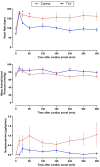
Figure 2. Hemodynamic parameters.
Heart rate (top), mean blood pressure (middle), and doses of norepinephrine administered after resuscitation (bottom) to prevent hypotension in animals subjected to the control procedure or to rapid cooling with TLV. *P<0.05 between the TLV and control group. TLV indicates total liquid ventilation.
Figure 3
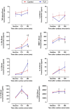
Figure 3. Blood circulatory inflammatory markers.
Blood concentration of cytokines at baseline, 120 minutes, and 360 minutes after cardiac arrest in animals subjected to the control procedure or to rapid cooling with TLV. *P<0.05 vs corresponding baseline. P<0.05 vs corresponding value at 120 minutes. CD31 indicates cluster of differentiation 31; IL, interleukin; MMP1, matrix metalloproteinase‐1; TLV, total liquid ventilation; and TNF‐alpha, tumor necrosis factor‐α.
Figure 4
Figure 4. Inflammatory markers in the CSF.
Cytokine concentrations in the CSF at 120 minutes and 360 minutes after cardiac arrest in animals subjected to the control procedure or to rapid cooling with TLV as compared with sham animals neither subjected to cardiac arrest nor TLV. *P<0.05 vs control group. P<0.05 vs sham. CD31 indicates cluster of differentiation 31; CSF, cerebrospinal fluid; IL, interleukin; MMP1, matrix metalloproteinase‐1; TLV, total liquid ventilation; and TNF‐alpha, tumor necrosis factor‐α.
Figure 5
Figure 5. Brain injury markers at the end of the protocol.
A, NFL blood levels at baseline and 360 minutes after cardiac arrest in animals subjected to the control procedure or to rapid cooling with TLV. B, Mean number of degenerating neurons spotted with Fluoro‐Jade C staining per field in the cortex. C, Morphological appearance of the cortex after Fluoro‐Jade C staining with few positive cells in the TLV vs control groups D, Mean number of cluster de différenciation 3 positive (CD3+) cells per field in the cortex, allowing to identify infiltrated T cells into the brain. E, Morphological appearance of the cortex after immunohistochemistry staining showing no or few CD3+ cells in the TLV vs control group. *P<0.05 vs control. P<0.05 vs corresponding baseline. NFL indicates neurofilament light chain; and TLV, total liquid ventilation.
Figure 6
Figure 6. Histological appearance of the lung.
A, Lesional scores on a 0 to 5 scale for cardiogenic serous edema, lung congestion, interstitial inflammation, and infarct‐like lesions of pulmonary parenchyma. B, Morphological appearance of a lung sample from the control group demonstrating extensive and severe serous edema and congestion. C, Morphological appearance of a lung sample from the TLV group demonstrating moderate congestion without alveolar serous edema. D, Morphological appearance of a lung sample from the control group demonstrating infarct‐like lesions. E, Morphological appearance of a lung sample from the TLV group with 1 macrophage with a foamy appearance (arrow). TLV indicates total liquid ventilation.

References

    1. Nolan JP, Neumar RW, Adrie C, Aibiki M, Berg RA, Böttiger BW, Callaway C, Rsb C, Geocadin RG, Jauch EC, et al. Post‐cardiac arrest syndrome: epidemiology, pathophysiology, treatment, and prognostication. A scientific statement from the International Liaison Committee on Resuscitation; the American Heart Association Emergency Cardiovascular Care Committee; the Council on Cardiovascular Surgery and Anesthesia; the Council on Cardiopulmonary, Perioperative, and Critical Care; the Council on Clinical Cardiology; the Council on Stroke. Resuscitation. 2008;79:350–379. doi: 10.1016/j.resuscitation.2008.09.017 - DOI - PubMed
    1. Perkins GD, Callaway CW, Haywood K, Neumar RW, Lilja G, Rowland MJ, Sawyer KN, Skrifvars MB, Nolan JP. Brain injury after cardiac arrest. Lancet Lond Engl. 2021;398:1269–1278. doi: 10.1016/S0140-6736(21)00953-3 - DOI - PubMed
    1. Logue ES, McMichael MJ, Callaway CW. Comparison of the effects of hypothermia at 33°C or 35°C after cardiac arrest in rats. Acad Emerg Med. 2007;14:293–300. doi: 10.1197/j.aem.2006.10.097 - DOI - PubMed
    1. Chenoune M, Lidouren F, Adam C, Pons S, Darbera L, Bruneval P, Ghaleh B, Zini R, Dubois‐Randé J‐L, Carli P, et al. Ultrafast and whole‐body cooling with total liquid ventilation induces favorable neurological and cardiac outcomes after cardiac arrest in rabbits. Circulation. 2011;124:901–911. doi: 10.1161/CIRCULATIONAHA.111.039388 - DOI - PMC - PubMed
    1. Nielsen N, Wetterslev J, Cronberg T, Erlinge D, Gasche Y, Hassager C, Horn J, Hovdenes J, Kjaergaard J, Kuiper M, et al. Targeted temperature management at 33°C versus 36°C after cardiac arrest. N Engl J Med. 2013;369:2197–2206. doi: 10.1056/NEJMoa1310519 - DOI - PubMed

LinkOut - more resources