. 2024 Oct;11(39):e2408024.
doi: 10.1002/advs.202408024. Epub 2024 Aug 19.

Ubiquitination of NS1 Confers Differential Adaptation of Zika Virus in Mammalian Hosts and Mosquito Vectors

Affiliations

Ubiquitination of NS1 Confers Differential Adaptation of Zika Virus in Mammalian Hosts and Mosquito Vectors

Chenxiao Huang et al. Adv Sci (Weinh). 2024 Oct.

Abstract

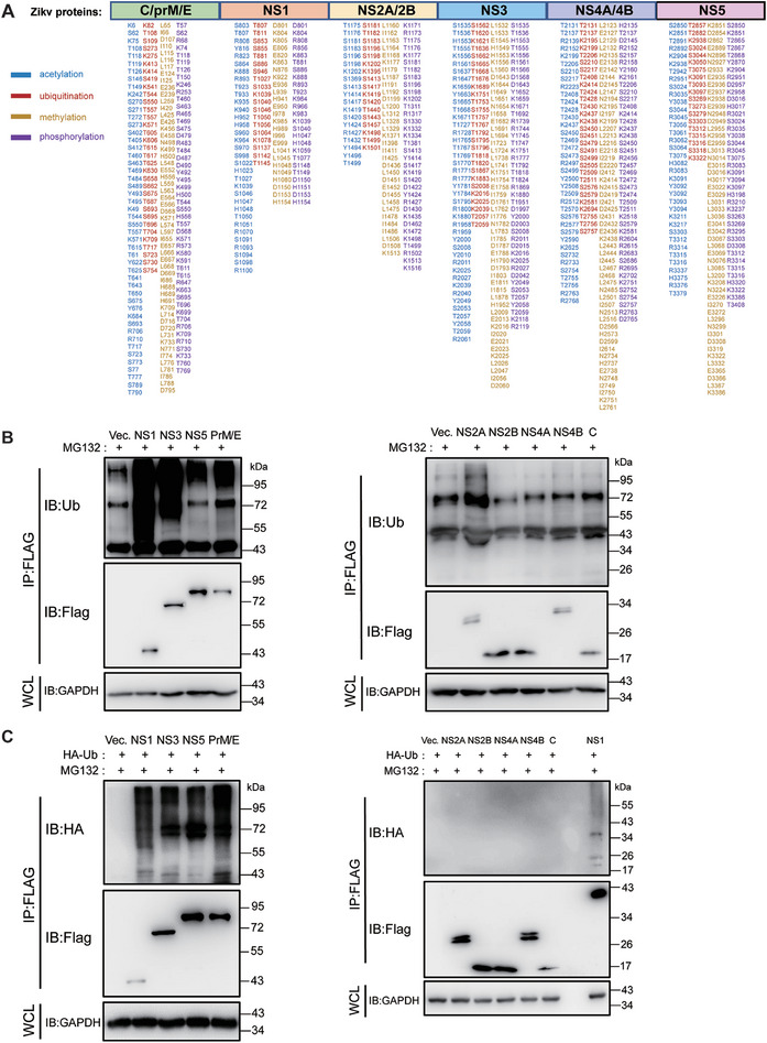
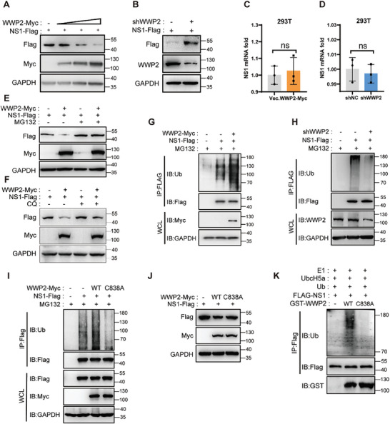
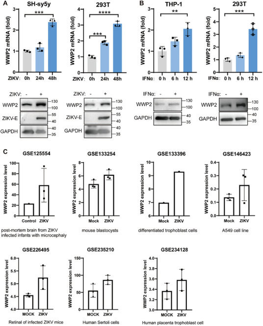
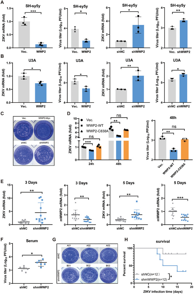
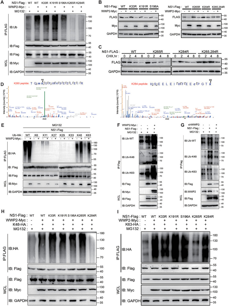
Arboviruses, transmitted by medical arthropods, pose a serious health threat worldwide. During viral infection, Post Translational Modifications (PTMs) are present on both host and viral proteins, regulating multiple processes of the viral lifecycle. In this study, a mammalian E3 ubiquitin ligase WWP2 (WW domain containing E3 ubiquitin ligase 2) is identified, which interacts with the NS1 protein of Zika virus (ZIKV) and mediates K63 and K48 ubiquitination of Lys 265 and Lys 284, respectively. WWP2-mediated NS1 ubiquitination leads to NS1 degradation via the ubiquitin-proteasome pathway, thereby inhibiting ZIKV infection in mammalian hosts. Simultaneously, it is found Su(dx), a protein highly homologous to host WWP2 in mosquitoes, is capable of ubiquitinating NS1 in mosquito cells. Unexpectedly, ubiquitination of NS1 in mosquitoes does not lead to NS1 degradation; instead, it promotes viral infection in mosquitoes. Correspondingly, the NS1 K265R mutant virus is less infectious to mosquitoes than the wild-type (WT) virus. The above results suggest that the ubiquitination of the NS1 protein confers different adaptations of ZIKV to hosts and vectors, and more importantly, this explains why NS1 K265-type strains have become predominantly endemic in nature. This study highlights the potential application in antiviral drug and vaccine development by targeting viral proteins' PTMs.

Keywords: E3 ligase WWP2; NS1; Zika virus; flavivirus; mosquito; ubiquitination.


Conflict of interest statement

The authors declare no conflict of interest.

Figures

Figure 1
Figure 1
Post‐translational modification of ZIKV viral proteins. A) SH‐sy5y cells were infected with ZIKV (MR766) at a multiplicity of infection (MOI) of 1, and cell samples were collected 48 h post‐infection. Mass spectrometry was employed to analyze the acetylation, methylation, phosphorylation, and ubiquitination patterns of viral proteins. B and C) 293T cells were transfected with plasmids encoding various ZIKV proteins tagged with Flag (NS1‐Flag, NS3‐Flag, NS5‐Flag, PrM/E‐Flag, NS2A‐Flag, NS2B‐Flag, NS4A‐Flag, NS4B‐Flag, or C‐Flag). B) In another set of experiments, these proteins were co‐transfected with a plasmid expressing ubiquitin tagged with HA (Ub‐HA). C) After 24 h, cells were treated with MG132 (5 µM) for 4 h. Cell lysates were collected, and immunoprecipitation with Flag antibody‐coupled magnetic beads was performed. The ubiquitination levels of viral proteins were then analyzed by Western Blot. The presented data are representative of three independent experiments.
Figure 2
Figure 2
E3 ubiquitin ligase WWP2 interacts with NS1. A) NS1‐Flag was transfected into 293T cells, followed by ZIKV infection (MOI = 0.5) 24 h later. Immunoprecipitation of NS1‐Flag was performed with Flag antibody‐coupled magnetic beads 24 h post‐infection to analyze the ubiquitin‐associated enzymes interacting with NS1‐Flag protein using mass spectrometry. B–E) WWP2‐Myc was co‐transfected with NS1‐Flag expression plasmid in 293T cells. After 24 h, immunoprecipitation of NS1‐Flag was performed, and WWP2‐Myc protein was detected by Western Blot. B) In another set of experiments, immunoprecipitation of WWP2‐Myc was carried out, and NS1‐Flag protein was detected by Western Blot. C) In addition, 293T cells were infected with ZIKV (MOI = 1), and after 48 h, endogenous WWP2 was immunoprecipitated. ZIKV NS1 protein was then detected by Western Blot D), and endogenous WWP2 protein was detected by immunoprecipitation of ZIKV NS1 and Western Blot E). F) 293T cells were infected with ZIKV(MOI = 0.5) for 48 h. The intracellular localization of the endogenous WWP2 and NS1 proteins was observed using laser confocal imaging. G and H) Each of the WWP2 truncates was co‐transfected with NS1‐Flag in 293T cells, and the cells were collected after 24 h. Immunoprecipitation with NS1‐Flag was performed, and Western Blot detected the expression of Myc‐tagged truncated proteins. I) Molecular docking prediction results for WWP2‐WW and ZIKV NS1 proteins. The data presented are representative of three independent experiments.
Figure 3
Figure 3
WWP2 ubiquitinates NS1 and leads to NS1 degradation. A and B) NS1‐Flag and WWP2‐Myc/shWWP2 (1 µg) were co‐transfected in 293T cells, and the protein levels of NS1‐Flag were detected by Western Blot 48 h later. C and D) NS1‐Flag and WWP2‐Myc/shWWP2 (1 µg) were co‐transfected in 293T cells, and the mRNA levels of NS1 were detected by qRT‐PCR 48 h later. E) NS1‐Flag and WWP2‐Myc were co‐transfected in 293T cells, which were then treated with MG132 (5 µM, 4 h) 24 h later. The protein levels of NS1‐Flag were detected by the Western Blot method. F) NS1‐Flag and WWP2‐Myc were co‐transfected in 293T cells. 24 h later, cells were treated with Chloroquine (10 µM, 6 h). The protein levels of NS1‐Flag were detected by the Western Blot. G and H) NS1‐Flag and shWWP2/WWP2‐Myc (1 µg) were co‐transfected in 293T cells and treated with MG132 (5 µM) for 4 h after 48 h. The ubiquitination level of NS1‐Flag protein was detected by the Western Blot method after immunoprecipitation of NS1‐Flag. I and J) NS1‐Flag and WWP2‐Myc (WT or C838A) plasmids were co‐transfected in 293T cells. After 24 h, immunoprecipitation of NS1‐Flag was performed, and the ubiquitination levels of NS1‐Flag protein were detected by Western Blot (F). The protein levels of NS1‐Flag were detected by Western Blot (G). K) NS1‐Flag, purified WWP2 (or WWP2‐C838A), E1 (Hdm2), and E2 (UbcH5a) were incubated for 1 h in the presence of ATP. The in vitro ubiquitination level of NS1 was analyzed by Western Blot. The data presented are representative of three independent experiments. ns, non‐significant (Student's t‐test).
Figure 4
Figure 4
WWP2 expression was upregulated during ZIKV infection. A) SH‐sy5y or 293T cells were infected with ZIKV (MR766) (MOI = 0.5). WWP2 levels was analyzed by qRT‐PCR and Western Blot. B) THP‐1 or 293T cells were treated with IFN‐α (500 U ml−1) for 6 h, and WWP2 levels were determined using qRT‐PCR and Western Blot. C) WWP2 expression was up‐regulated during ZIKV infection based on GEO databases. Data are representative of 3 independent experiments and presented as mean ± SD. ** P < 0.01, and *** P < 0.001, **** P < 0.0001 (Student's t‐test).
Figure 5
Figure 5
WWP2 restricts ZIKV infection. A–C) SH‐sy5y (A)/U3A (B) cells were infected with lentivirus overexpressing or knocking down WWP2 (MOI = 10). Subsequently, cells were infected with ZIKV (MOI = 0.5) 48 h later. Viral mRNA levels in the cells were detected 24 h later using qRT‐PCR. Viral load in the supernatant was visualized by TCID50, and the infectious viral load in U3A supernatants was determined by plaque assay (C). D) WWP2‐Myc (WT or C838A) plasmid was transfected into 293T cells and infected with ZIKV (MOI = 0.5) after 24 h. Cellular RNA was extracted at 24 and 48 h, and the viral RNA levels were analyzed by qRT‐PCR. The viral supernatant titer after 48 h was determined by TCID50. E‐H) Ifnar1−/− mice (6 weeks old, 12 mice per group) were injected with 5 × 107 PFU shmWWP2 lentivirus via the tail‐vein route; 7 days later, mice were injected intraperitoneally with 107 PFU ZIKV. Hemocytes and serum were collected on days 3 and 5. Blood cell RNA was extracted, and qRT‐PCR was used to detect the RNA content of ZIKV and shmWWP2 in the cells E). Viral titers in the serum of mice on day 5 were detected by TCID50 F). Infectious virus in the serum of mice on day 5 was detected by the plaque assay G). The status and survival of mice were recorded by daily observation (* P < 0.05, Log‐rank test) (H). Data are representative of 3 independent experiments and presented as mean ± SD. ns, non‐significant, * P < 0.05, ** P < 0.01, and *** P < 0.001 (Student's t‐test).
Figure 6
Figure 6
WWP2 ubiquitinates amino acids K265 and K284 of ZIKV NS1. A and B) WWP2‐Myc was co‐transfected with NS1‐WT or its mutants in 293T cells for 24 h. After 24 h, the cells were treated with MG132 (5 µM) for 4 h. NS1‐Flag was immunoprecipitated, and the ubiquitylation level of NS1 proteins was detected by Western Blot A). The protein level of NS1 was detected by Western Blot B). C) NS1‐WT or its mutants were transfected into 293T cells and treated with actinomycin ketone CHX (50 µM) for 0, 2, 4, and 8 h after 24 h. NS1 protein levels were detected by Western Blot. D) Secondary mass spectrometry analysis of ubiquitinations at positions K265 and K284 of NS1. E) WWP2‐Myc, NS1‐Flag, and ubiquitin molecule mutant plasmids (K6, K11, K27, K29, K33, K48, and K63) were co‐transfected into 293T cells. After 24 h, the cells were treated with MG132 (5 µM, 4 h), and NS1‐Flag was immunoprecipitated. NS1 proteins were detected by Western Blot method, and the ubiquitination level was assessed. F and G) WWP2‐Myc or shWWP2 (1 µg), NS1‐Flag, were co‐transfected into 293T cells. After 24 h, the cells were treated with MG132 (5 µM, 4 h), and NS1‐Flag was immunoprecipitated. NS1 proteins were detected by Western Blot, and the ubiquitination types of NS1 were detected using K48 and K63 antibodies. H and I) WWP2‐Myc, NS1‐Flag individual point mutants, and ubiquitin molecule mutant K48/K63‐HA were co‐transfected into 293T cells. After 24 h, the cells were treated with MG132 (5 µM, 4 h), and immunoprecipitated with NS1‐Flag. The level of ubiquitination of NS1 proteins was detected by Western Blot. Data are representative of 3 independent experiments.
Figure 7
Figure 7
Amino acid mutation at position NS1 K265, K284 alters ZIKV virulence. A and B) Schematic diagram of the ZIKV packaging process (A): The full‐length plasmid of the 2016 GZ01 strain was used as a template. The full‐length plasmid of the NS1 point‐mutated K265R, K284R, and K265/284R viral genomes was obtained by targeted mutagenesis. The full‐length plasmid was transfected with RNA into BHK21 cells after in vitro transcription, and the viral supernatant was collected after culture to obtain the WT viruses and mutant viruses (B). C–E) WT, K265R, K284R, and K265/284R viruses were packaged with the same mass of RNA, and the titer of the viral particles was detected by TCID50 (C). 293T cells were infected with the same titer of the mutant viruses (MOI = 1), and the intracellular viral load was detected by qRT‐PCR 48 h later (D). The same titer of mutant viruses was used to infect 293T cells (MOI = 1), and the level of ZIKV NS1 in the supernatant was detected by ELISA after 72 h (E). F and G) 293T (F) and SH‐sy5y (G) cells were infected with lentiviruses knocking down the expression of WWP2, and then infected with WT and mutant viruses after 48 h. Cells were collected after 48 h to extract the RNA, and the viral load in the cells was detected by qRT‐PCR. H) Ifnar1−/− mice were infected with 107 PFU viruses (WT, K265R, K284R, and K265/284R), and viral loads were detected by qRT‐PCR on day 5 after infection. I) A search of the Virus Sequence Library (https://nextstrain.org) revealed the existence of a naturally occurring strain of ZIKV NS1 mutated at amino acid positions 265/284. J) Model for regulation of ZIKV NS1 by WWP2. Data are representative of 3 independent experiments and presented as mean ± SD. ns, non‐significant, * P < 0.05, ** P < 0.01, and *** P < 0.001, **** P < 0.0001 (Student's t‐test).
Figure 8
Figure 8
WWP2 is a broad‐spectrum arthropod‐borne flavivirus suppressor. A) Conservation of site 284 of the arthropod‐borne flavivirus NS1 protein. B–D) After overexpression or knockdown of WWP2 in 293T (B) and U3A (C) cells, the cells were infected with JEV (MOI = 0.5), and cellular RNA was extracted after 24 and 48 h. The viral content of the cells was detected by qRT‐PCR; the amount of infectious viruses in the supernatant of U3A was detected by plaque assay (D). E–G) Using WT and Wwp 2−/− mice, 107 PFU JEV (SA14) was injected intraperitoneally, and hemocytes and serum were collected by orbital blood sampling on days 3 and 5, respectively. Blood cell RNA was extracted, and the amount of JEV in the cells was detected using qRT‐PCR (E); the viral titer in the serum of mice on day 5 was detected by TCID50 (F); the survival of mice was observed and recorded daily (* P<0.05, Log‐rank test) (G). H) JEV NS1‐Flag and WWP2‐Myc/shWWP2 (1 µg) plasmids were co‐transfected in 293T cells and treated with MG132 (5 µM) for 4 h after 48 h. NS1‐Flag was immunoprecipitated, and ubiquitination of JEV NS1‐Flag protein was detected by Western Blot. I) JEV NS1‐Flag and WWP2‐Myc/shWWP2 (1 µg) plasmids were co‐transfected in 293T cells, and the cells were collected after 48 h. NS1 protein levels were detected by Western Blot. J) JEV NS1‐Flag and WWP2‐Myc were co‐transfected in 293T cells, which were treated with MG132 (5 µM) for 4 h after 24 h. JEV NS1 protein levels were detected by Western Blot. K‐N) Cells were infected with LGTV after overexpression or knockdown of WWP2 in 293T (K) and U3A cells (M) (MOI = 1), and cellular RNA was extracted after 48 h. The viral RNA load in the cells was detected by using qRT‐PCR; etch‐a‐sketch assay was performed to detect the amount of infectious virus in the supernatants of U3A cells (N). Data are representative of 3 independent experiments and presented as mean ± SD. *P < 0.05, **P < 0.01, and ***P < 0.001 (Student's t‐test).
Figure 9
Figure 9
Ubiquitination of NS1 by WWP2 homologs in mosquitoes promotes ZIKV infection of mosquitoes. A) ZIKV NS1‐Flag was transfected in C6/36 cells and treated with MG132 (5 µM) for 4 h after 24 h. Cells were collected and immunoprecipitated with Flag antibody‐coupled magnetic beads, and the ubiquitination level of viral proteins was detected by Western Blot. B) The E3 ligase Su(dx), which is highly homologous to human WWP2, is present in Aedes albopictus. (WWP2 GenBank: U96114.2; Su(dx) GenBank: XM_01 969 6185.2) C) NS1‐Flag was co‐transfected with Su(dx)‐His expression plasmid in C6/36 cells and infected with ZIKV (MR766) (MOI = 0.5) 24 h later for 24 h. NS1‐Flag was immunoprecipitated, and Su(dx)‐His protein was detected by Western Blot. D) NS1‐Flag and different doses of Su(dx) were co‐transfected in C6/36 cells, and NS1‐Flag protein levels were detected by Western Blot 24 h later. E) Co‐transfected siSu(dx) (50 nM) with NS1‐Flag (1 µg) in C6/36 cells, treated with MG132 (5 µM) for 4 h after 24 h. Immunoprecipitation of NS1‐Flag was performed, and the ubiquitination level of NS1 protein was detected by Western Blot method. F) Su(dx)‐Flag (1 µg) was transfected into C6/36 cells. After 24 h, the cells were infected with WT, K265R, K284R and K265, 284R viruses (MOI = 0.5), respectively. 48 h later, the cells were treated with MG132 (5 µM, 4 h) and NS1 was immunoprecipitated. NS1 protein was detected by Western Blot and its ubiquitination level was determined. G) Transfection of siSu(dx) (50 nM) in C6/36 cells was followed by infection with ZIKV (MOI = 1) after 48 h. Viral mRNA levels in the cells, as well as Su(dx) knockdown efficiency, were detected after 24 h using qRT‐PCR. H) Su(dx)‐His was transfected in C6/36 cells, infected with ZIKV (MOI = 1) 24 h later, and the viral mRNA level as well as the efficiency of Su(dx) overexpression was detected in the cells 48 h later using qRT‐PCR. I) NSC2805 (10 µM, 4 h) treated C6/36 cells were infected with ZIKV and viral RNA levels were detected by qRT‐PCR at 24 h J) Aedes aegypti mosquitoes were divided into two groups, the experimental group was injected with Su(dx) dsRNA, and the control group was injected with Luc dsRNA. 100 PFU of MR766 strain virus was injected into each mosquito. The viral mRNA level and Su(dx) knockdown efficiency in mosquitoes were detected by qRT‐PCR on day 7 after infection. K) Recombinant viruses (WT, K265R, K284R, and K265/284R) of the same titer were injected into the thoracic cavity of Aedes aegypti mosquitoes (50 PFU of virus per mosquito), and viral loads in the mosquitoes were detected by qRT‐PCR on day 7 after infection. Data are representative of 3 independent experiments and presented as mean ± SD. ns, non‐significant, * P < 0.05, **P < 0.01, and ***P < 0.001, ****P < 0.0001 (Student's t‐test).

Similar articles

Cited by

References

    1. Wu P., Yu X., Wang P., Cheng G., Expert Rev. Mol. Med. 2019, 21, e1. - PubMed
    1. a) Collins M. H., Metz S. W., Clin. Ther. 2017, 39, 1519; - PubMed
    2. b) Scherwitzl I., Mongkolsapaja J., Screaton G., Current Opin. Virol. 2017, 23, 95. - PubMed
    1. Kumar R., Mehta D., Mishra N., Nayak D., Sunil S., Int. J. Mol. Sci. 2020, 22. - PMC - PubMed
    1. a) Santos A. L., Lindner A. B., Oxid. Med. Cell Longev. 2017, 2017, 5716409; - PMC - PubMed
    2. b) Rahnefeld A., Klingel K., Schuermann A., Diny N. L., Althof N., Lindner A., Bleienheuft P., Savvatis K., Respondek D., Opitz E., Ketscher L., Sauter M., Seifert U., Tschöpe C., Poller W., Knobeloch K. P., Voigt A., Circulation 2014, 130, 1589; - PubMed
    3. c) Zhang T., Ye Z., Yang X., Qin Y., Hu Y., Tong X., Lai W., Ye X., Sci. Rep. 2017, 7, 43691. - PMC - PubMed
    1. Bharaj P., Atkins C., Luthra P., Giraldo M. I., Dawes B. E., Miorin L., Johnson J. R., Krogan N. J., Basler C. F., Freiberg A. N., Rajsbaum R., J. Virol. 2017, 91. - PMC - PubMed

MeSH terms

Substances

LinkOut - more resources