Review
. 2024 Oct;11(39):e2308900.
doi: 10.1002/advs.202308900. Epub 2024 Aug 19.

From Cell to Gene: Deciphering the Mechanism of Heart Failure With Single-Cell Sequencing

Affiliations
Review

From Cell to Gene: Deciphering the Mechanism of Heart Failure With Single-Cell Sequencing

Dan Zhang et al. Adv Sci (Weinh). 2024 Oct.

Abstract

Heart failure (HF) is a prevalent cardiovascular disease with significant morbidity and mortality rates worldwide. Due to the intricate structure of the heart, diverse cell types, and the complex pathogenesis of HF, further in-depth investigation into the underlying mechanisms is required. The elucidation of the heterogeneity of cardiomyocytes and the intercellular communication network is particularly important. Traditional high-throughput sequencing methods provide an average measure of gene expression, failing to capture the "heterogeneity" between cells and impacting the accuracy of gene function knowledge. In contrast, single-cell sequencing techniques allow for the amplification of the entire genome or transcriptome at the individual cell level, facilitating the examination of gene structure and expression with unparalleled precision. This approach offers valuable insights into disease mechanisms, enabling the identification of changes in cellular components and gene expressions during hypertrophy associated with HF. Moreover, it reveals distinct cell populations and their unique roles in the HF microenvironment, providing a comprehensive understanding of the cellular landscape that underpins HF pathogenesis. This review focuses on the insights provided by single-cell sequencing techniques into the mechanisms underlying HF and discusses the challenges encountered in current cardiovascular research.

Keywords: application; challenging; heart failure; mechanism; single‐cell sequencing.


Conflict of interest statement

The authors declare no conflict of interest.

Figures

Figure 1
Figure 1
The basic flow of single‐cell sequencing. Single‐cell isolation is the process of obtaining single cells from heart tissue using multiple methods, including the gradient dilution method, negative pressure suction method, micromanipulation method, fluorescence‐activated cell sorting (FACS), laser capture microdissection (LCM), microarray technology, microfluidic techniques, and droplet technology, and the ICELL8 cx Single‐Cell System. Single‐cell amplification refers to increasing the amount of genetic material, either DNA or RNA, from a single cell to enable further analysis. Single‐cell DNA amplification replicates the genomic DNA of a single cell, while single‐cell RNA amplification replicates the RNA transcripts present in a single cell. Library construction for scRNA‐seq involves converting RNA into complementary DNA (cDNA), adding adapters, and amplifying the cDNA. The sequencing step then determines the order of nucleotides in the cDNA. Data analysis for sscRNA‐seq involves processing and interpreting the sequencing data to identify and characterize gene expression patterns at the individual cell level. This analysis includes steps such as quality control, normalization, dimensionality reduction, clustering, differential gene expression analysis, and functional annotation to gain insights into the cell populations and their biological functions.
Figure 2
Figure 2
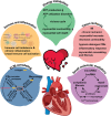
Application of SCS in studying the mechanisms of HF. With extensive use of SCS in cardiovascular, the understanding of the mechanisms of HF has deepened, mainly manifested in the following aspects: 1. energy metabolism, 2. immune and inflammation, 3. tissue ischemia, 4. ECM remodling, 5. small molecular mediators. Studying the structure and function of the heart at the single‐cell level provides reliable ideas for the study of the molecular mechanism of HF and the further treatment of heart diseases. However, we must also meet more challenges and problems with the mechanism of HF based on single‐cell studies.
Figure 3
Figure 3
Impaired myocardial energy metabolism mediates the onset and progression of HF in several ways. A) The gene expression variability in CMs after HF. B) Cardiac morphological hypertrophy is associated with enhanced mitochondrial biosynthesis. C) The impaired intracellular metabolism of CMs in HF promotes the entry of lipid molecules into lipotoxic pathways/signaling, which further induces possible cardiac dysfunction. D) The expression of energy metabolism‐related genes mainly includes FA oxidation, TCA, and glycolysis FA uptake, which initially increases and then decreases with TAC over time. E) Mitochondrial damage leads to ATP reduction, and conversely, ATP reduction will also cause mitochondrial damage, resulting in myocardial cell death, cardiac fibrosis, and HF (Figure 3E). CMs, cardiomyocytes; TAC, transverse aorta constriction; PE, phosphatidylethanolamines; PI, phosphatidylinositol; PC, phosphatidylcholines. FA, fatty acids; FAO, fatty acids oxidation; TCA, tricarboxylic acid; OXPHOS, oxidative phosphorylation; mt, mitochondrion; ATP, adenosine 5′‐triphosphate; ECC, excitation‐contraction coupling; BACCs, branch‐chain amino acids.
Figure 4
Figure 4
Immune cell imbalance and inflammation promote the progression of HF. The involvement of both the innate and adaptive immune systems in HF leads to localized immune responses in the heart, contributing to ventricular remodeling and dysfunction. Various types of immune response cells play a significant role in the development of HF. These cells are activated, transitioned, and regulate CMs, ECs, and FBs through complex immune and inflammatory processes during the progression of the disease. ACKR1, atypical chemokine receptor 1; ACTA2, actin alpha 2; AEBP1, AE Binding Protein 1; B, B cells; CMs, cardiomyocytes; CCR2, CC chemokine receptor 2; CSF1R, colony‐stimulating factor 1 receptor; CXCL8, C‐X‐C motif chemokine ligand 8 (interleukin 8); DAMPs, damage‐associated molecular patterns; DARC, Duffy antigen receptor for chemokines; ECs, endothelial cells; FBs, fibroblasts; HF, heart failure; HTRA3, HtrA serine peptidase 3; IGFBP7, insulin‐like growth factor‐binding protein‐7; NK, natural killer cells; OSM, cytokine Oncostatin M; PD‐1, programmed cell death protein 1; T, T cells; TGF‐β, transforming growth factor‐beta.
Figure 5
Figure 5
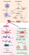
Small molecular mediators in heart failure indicated by SCS. Pressure overload induces miR‐21 increase in cardiac macrophage, which can activate FBs through paracrine. But ischania/hypoxia induces miR‐51 decrease, inhibiting FBs transformation. Both of them can induce fibrosis and promote HF progress. MiR‐51 also upregulates SPRR1A in ischemic HF, which can suppress fibrosis. LncRNA also are involved in HF. Knockout of Trdn‐as damages the recruitment of splicing factors to Trdn pre‐mRNA, causing abnormal composition of triadin isoforms in the heart, Ca2+ mishandling, and susceptibility to cardiac arrhythmias. LncRNAs Gas5 and Sghrt in FBs are upregulated after stress response or MI, regulating co‐expressed genes within the same gene regulatory network including cell cycle genes: CCNG1 and CCND2 and others: NPPA and DSTN. AS, alternative splice; ECM, extracellular matrix; HF, heart failure; KO, Knockout; FBs, fibroblasts; MI, myocardial infarction; lncRNA, long noncoding RNA; Trdn‐as, a CM‐specific lncRNA encoded by the antisense strand of the triadin gene.
Figure 6
Figure 6
Inter‐cellular communication in the heart as a possible therapeutic target for HF. Extensive cellular interactions between different types of cardiac cells play an important role in HF. Inter‐cellular communication in the heart mainly includes paracrine signaling and autocrine signaling. Macrophages communicate with CMs, ECs, and FBs through receptor‐ligand interaction, such as C5AR1 in macrophages as a receptor interacting with RPS19 in CMs, ECs, and FBs as a ligand for paracrine signaling. FBs express CSF1 and IL34, which regulate macrophage growth and survival through CSF1R signaling. FBs also express growth factors, including NGF, VEGFA, IGF1, and FGF2, which are vital for supporting the development of neurons in the autonomic nervous system, ECs, and parietal cells. CMs, cardiomyocytes; ECs, endothelial cells; ESAM, endothelial cell‐selective adhesion molecule; FBs, fibroblasts; NGF, nerve growth factor; VEGFA, vascular endothelial growth factor A; IGF1, insulin‐like growth factor‐1; FGF2, fibroblast growth factor 2.
Figure 7
Figure 7
From cell to gene: multiple mechanisms work together to mediate HF. Cell composition and cell types undergo extensive changes during HF. ECs, FBs, and immune cells all contribute to the occurrence of HF. ECs affect myocardial fibrosis mainly through regulating EndMT and paracrine signaling pathways, such as SIRT1 activated by resveratrol attenuated cardiac fibrosis by regulating EndMT via the TGF‐β/Smad2/3 pathway. FBs in HF and cardiac fibrosis also induced ECs inflammation and dysfunction in a paracrine manner. Immune cells, such as macrophages, T cells, neutrophils, B cells, NK cells, and mast cells, are extensively activated after pressure overload and are involved in HF progression. AEBP1, AE Binding Protein 1; B, B cells; CAMs, cell adhesion molecule; CCR2, CC chemokine receptor 2; CKAP4, cytoskeleton‐associated protein 4; CSF1R, colony stimulating factor 1 receptor; D1R, dopamine D1 receptor; ECM, extracellular matrix; EndMT, endothelial‐to‐mesenchymal transition; FBs, fibroblasts; HTRA3, HtrA serine peptidase 3; M1‐like, M1‐like macrophage; NOX4, NADPH oxidase 4; OSM, cytokine oncostatin M; PD‐1, programmed cell death protein 1; RyR2, ryanodine receptor 2; ROS, reactive oxygen species; TAC, transverse aortic constriction; TEAD1, TEA domain transcription factor 1; TGF‐β, transforming growth factor‐beta; TF, transcription factor.

References

    1. Tang F., Barbacioru C., Wang Y., Nordman E., Lee C., Xu N., Wang X., Bodeau J., Tuch B. B., Siddiqui A., Lao K., Surani M. A., Nat. Methods 2009, 6, 377. - PubMed
    1. Ishii S., Tago K., Senoo K., Appl. Microbiol. Biotechnol. 2010, 86, 1281. - PubMed
    1. Zhu Z., Chen X., Genom. Appl. Biol. 2015, 34, 902.
    1. Yan D., C. Song, He H., Chem. Indust. Engineer. 2015, 32, 71.
    1. Lu Z., Moraes C., Zhao Y., You L., Simmons C. A., Sun Y., in IEEE International Conference on Robotics and Automation 2010, 494.

LinkOut - more resources