. 2024 Aug 20;150(8):657-660.
doi: 10.1161/CIRCULATIONAHA.123.064343. Epub 2024 Aug 19.

Junctophilin-2 Regulates Mitochondrial Metabolism

Affiliations

Junctophilin-2 Regulates Mitochondrial Metabolism

Sasha Z Prisco et al. Circulation. .
No abstract available

Keywords: metabolism; mitochondria; ventricular dysfunction, right.


Conflict of interest statement

Dr Prins obtained funding from Bayer to support this work. The other authors have no relevant disclosures.

Figures
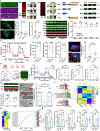
Figure:
Figure:. JPH2 binds MFN2 and regulates mitochondrial metabolism and RV function.
(A) Confocal micrographs demonstrated JPH2 (green) and MFN2 (purple) co-localized in isolated RV cardiomyocytes. Coomassie brilliant blue (CBB) of SDS-PAGE of cytoplasmic and mitochondrial extracts. Western blot analysis of cellular fractionations showed both JPH2 and MFN2 were enriched in the mitochondrial fraction. Co-immunoprecipitation studies demonstrated a biochemical interaction between JPH2 and MFN2. Immunoblots of pulldown experiments of recombinantly expressed and purified JPH2 and MFN2 constructs revealed the two proteins directly bound each other, and the interaction site was localized to the amino-terminal third of JPH2. (B) Representative confocal micrographs revealed disruption of mitochondrial network morphology (p-values determined by Mann-Whitney U-test) in JPH2 KO iPSC-CM. Western blot analysis showed JPH2 expression was abolished in JPH2 KO iPSC-CM (above) without significantly altering mitochondrial fission/fusion protein expression (p-values determined by unpaired t-test). CBB gel showed equivalent loading (below). (C) Seahorse tracings and quantification of reduced oxygen consumption rates (OCR) in JPH2 KO iPSC-CM (p-values determined by unpaired t-test). Confocal micrographs of lipid droplets (left) and the area of cells occupied by lipid droplets in control and JPH2 KO iPSC-CM. (D) Re-expression of full-length JPH2 (JPH2FL) augmented mitochondrial oxygen consumption as compared to C-terminal JPH2 (JPH2Ct) overexpression (p-values determined by one-way ANOVA with Tukey’s multiple comparisons test or by Kruskal-Wallis test with Dunn’s multiple comparisons test). (E) AAV-JPH2 restored JPH2 protein abundance and did not significantly impact MFN2 levels (p-values determined by one-way ANOVA with Tukey’s multiple comparisons test) and improved peri-t-tubular (red arrow) mitochondrial cristae structure (p-values determined by Kruskal-Wallis test with Dunn’s multiple comparisons test). (F) KEGG, Reactome, and WikiPathway analysis of differentially expressed proteins in mitochondrial enrichments. Red boxes highlight FAO related pathways. Hierarchical cluster analysis demonstrated AAV-JPH2 increased mitochondrial FAO protein abundance in RV mitochondrial extracts. (G) Hierarchical cluster and random forest analyses suggested AAV-JPH2 treatment restructured RV lipid homeostasis. (H) AAV-JPH2 treatment enhanced RV function as assessed by echocardiography and pressure-volume loop analysis without significantly altering RV afterload. p-values determined by one-way ANOVA with Tukey’s multiple comparisons test for TAPSE, RV free wall thickness change, RV-pulmonary artery coupling (Ees/Ea), and effective arterial elastance (Ea). p-values for RVSP determined by Brown-Forsythe ANOVA with Dunnett’s T3 multiple comparisons test.

Update of

References

    1. Prisco SZ, Thenappan T, Prins KW. Treatment Targets for Right Ventricular Dysfunction in Pulmonary Arterial Hypertension. JACC Basic Transl Sci. 2020;5:1244–1260. doi: 10.1016/j.jacbts.2020.07.011 - DOI - PMC - PubMed
    1. Lehnart SE, Wehrens XHT. The role of junctophilin proteins in cellular function. Physiol Rev. 2022;102:1211–1261. doi: 10.1152/physrev.00024.2021 - DOI - PMC - PubMed
    1. Takeshima H SK, Nishi M, Iino M, Kangawa K. Junctophilins: a novel family of junctional membrane complex proteins. In: Molecular Cell; 2000:11–22. doi: 10.1016/s1097-2765(00)00003-4 - DOI - PubMed
    1. Liang X, Mei Y, Huang X, Shen G, Zhu D, Yu Y, Wang J, Lou Y. Junctophilin 2 knockdown interfere with mitochondrium status in ESC-CMs and cardiogenesis of ES cells. J Cell Biochem. 2012;113:2884–2894. doi: 10.1002/jcb.24164 - DOI - PubMed
    1. Boucherat O, Yokokawa T, Krishna V, Kalyana-Sundaram S, Martineau S, Breuils-Bonnet S, Azhar N, Bonilla F, Gutstein D, Potus F, et al. Identification of LTBP-2 as a plasma biomarker for right ventricular dysfunction in human pulmonary arterial hypertension. Nature Cardiovascular Research. 2022;1:748–760. doi: 10.1038/s44161-022-00113-w - DOI

LinkOut - more resources