. 2024 Aug 19;20(8):e1011965.
doi: 10.1371/journal.ppat.1011965. eCollection 2024 Aug.

RNase-mediated reprogramming of Yersinia virulence

Affiliations

RNase-mediated reprogramming of Yersinia virulence

Ines Meyer et al. PLoS Pathog. .

Abstract

RNA degradation is an essential process that allows bacteria to regulate gene expression and has emerged as an important mechanism for controlling virulence. However, the individual contributions of RNases in this process are mostly unknown. Here, we tested the influence of 11 potential RNases in the intestinal pathogen Yersinia pseudotuberculosis on the expression of its type III secretion system (T3SS) and associated effectors (Yops) that are encoded on the Yersinia virulence plasmid. We found that exoribonuclease PNPase and endoribonuclease RNase III inhibit T3SS and yop gene transcription by repressing the synthesis of LcrF, the master activator of Yop-T3SS. Loss of both RNases led to an increase in lcrF mRNA levels. Our work indicates that PNPase exerts its influence via YopD, which accelerates lcrF mRNA degradation. Loss of RNase III, on the other hand, results in the downregulation of the CsrB and CsrC RNAs, thereby increasing the availability of active CsrA, which has been shown previously to enhance lcrF mRNA translation and stability. This CsrA-promoted increase of lcrF mRNA translation could be supported by other factors promoting the protein translation efficiency (e.g. IF-3, RimM, RsmG) that were also found to be repressed by RNase III. Transcriptomic profiling further revealed that Ysc-T3SS-mediated Yop secretion leads to global reprogramming of the Yersinia transcriptome with a massive shift of the expression from chromosomal to virulence plasmid-encoded genes. A similar reprogramming was also observed in the RNase III-deficient mutant under non-secretion conditions. Overall, our work revealed a complex control system where RNases orchestrate the expression of the T3SS/Yop machinery on multiple levels to antagonize phagocytic uptake and elimination by innate immune cells.


Conflict of interest statement

The authors have declared that no competing interests exist.

Figures
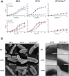
Fig 1
Fig 1. Loss of RNases PNPase and RNase III affects growth and the formation of T3S-like structures of Y. pseudotuberculosis.
(A) Overnight cultures of Y. pseudotuberculosis YPIII and the isogenic RNase mutants YP139 (Δpnp) and YP356 (Δrnc) harboring the empty vector (pV) or the complementation plasmids (prnc+ or ppnp+) were diluted to an OD600 of 0.2 in LB and growth at 25°C, 37°C, and 37°C/-Ca2+ was followed by measurement of OD600. Data represent the mean ± SD from experiments done in triplicates. (B) Scanning (left panel) and transmission (right panel) microscopy of Y. pseudotuberculosis strains YPIII (wt), YP139 (Δpnp), and YP356 (Δrnc) grown at 25°C, 37°C and 37°C/-Ca2+. Flagella are indicated by grey and T3S injectisome-like structures by white arrows, respectively. Bars indicate 200 nm.
Fig 2
Fig 2. Influence of PNPase and RNase III on Yop protein secretion.
(A) Y. pseudotuberculosis strains YPIII (wt), YP139 (Δpnp), and YP356 (Δrnc) were grown at 25°C, 37°C, and 37°C/-Ca2+; the secreted proteins in the supernatant of the cultures were precipitated with TCA and separated on SDS gels (left panels). The Yop secretion-deficient mutant YP101 (ΔyscS) was used as negative control. Secreted Yop proteins were quantified using ImageJ. Data represent the mean ± SD from three independent biological replicates relative to the amounts of Yops secreted by the wildtype at 25°C (right panel). Significant differences were determined using the one-way Anova test and are indicated by asterisks (*P <0.05, **P = 0.01). c: indicates an unidentified secreted protein used to control the efficiency of protein precipitation (B) Secretion of a plasmid-encoded YopE-beta-lactamase (BlaM) fusion protein (illustrated in the upper panel) expressed in Y. pseudotuberculosis strains YPIII (wt), and YP356 (Δrnc) grown at 37°C and 37°C/-Ca2+ for 4 h was determined. Data represent the mean ± SD from three independent biological replicates (lower panel). Significant differences were determined using the Student’s t-test and are indicated by asterisks (****P<0.0001).
Fig 3
Fig 3. Yop synthesis is altered in PNPase- and RNase III-deficient strains.
Y. pseudotuberculosis strains YPIII (wt), YP139 (Δpnp), and YP356 (Δrnc) without (A,B) or with a plasmid-encoded YopE-beta-lactamase (BlaM) fusion protein (C) were grown at 37°C (+) and 37°C/-Ca2+ (-); whole cell extracts were prepared and separated on SDS gels (left panels). Synthesized Yop proteins in the YPIII (wt) (A-C), YP139 (Δpnp) (A), YP356 (Δrnc) (B), and (C) YP356 (Δrnc) pIVO13 (yopE-blaM) were detected by Western blotting using a multi-Yop antiserum (left panel) and were quantified by ImageJ (right panel). An antiserum against H-NS was used for loading control. Data represent the mean ± SD from three independent biological replicates and relative amounts are documented with respect to the wildtype grown at 37°C. Significant differences were determined using Student’s t-test and indicated by asterisks (*P <0.05; ****P<0.0001).
Fig 4
Fig 4. Influence of RNase III and PNPase on the synthesis of LcrF.
Y. pseudotuberculosis strains YPIII (wt), YP139 (Δpnp) (A), and YP356 (Δrnc) (B) were grown at 37°C (+) and 37°C/-Ca2+ (-). The secretion-deficient mutant YP101 (ΔyscS) and the LcrF-deficient mutant strain YP179 (ΔlcrF) were used as a negative control. Whole-cell extracts were prepared and separated by SDS-PAGE. The transcriptional activator LcrF was detected by Western blotting using an LcrF-specific polyclonal antiserum. An antiserum against H-NS was used for loading control (left panel). Data were quantified by ImageJ and represent the mean ± SD from three independent biological replicates (right panel). Significant differences were determined using Student’s t-test and indicated by asterisks (*P <0.05; ***P<0.001). (C) Y. pseudotuberculosis strains YPIII (wt), YP139 (Δrnc), and YP356 (Δrnc) pIVO20 (prnc+) were grown at 37°C (+) and 37°C/-Ca2+ (-); the LcrF-deficient mutant strain YP179 was used as a negative control. Whole-cell extracts were prepared and separated on SDS gels. Synthesized LcrF was detected by Western blotting using a polyclonal LcrF antiserum. An antiserum against RNA polymerase (RNAP) was used for loading control.
Fig 5
Fig 5. Influence of RNase III and PNPase on lcrF transcript levels.
(A) Y. pseudotuberculosis strains YPIII (wt), YP139 (Δpnp), YP356 (Δrnc), and YP375 (Δrnc, Δpnp) were grown at 37°C and 37°C/-Ca2+. Total RNA of the samples was prepared and the lcrF transcript was detected by Northern blotting; 16S rRNA was used as loading control. (B) Y. pseudotuberculosis strains YPIII (wt), YP139 (Δpnp), YP356 (Δrnc), and YP356 (Δrnc) pIVO20 (prnc+) and the ΔlcrF strain (YP179) were grown at 37°C. Total RNA of the samples was prepared and the lcrF transcript was detected by Northern blotting (left panel) and quantified by ImageJ (right panel). Data represent the mean ± SD from three independent biological replicates. Significant differences were determined using Student’s t-test and indicated by asterisks (*P<0.05). (C) RNA-Seq coverage tracks of the yscW-lcrF locus in Y. pseudotuberculosis YPIII (wt) and YP356 (Δrnc) grown at 37°C. RNA-Seq reads were mapped to the pYV virulence plasmid (NC_006153.2) and analyzed using a DESeq2 pipeline. The mapping visualizes the nucleotide score of the combined biological replicates for the two strains with an equal score for both. (D) RNA-Seq coverage tracks of the yscW-lcrF locus of the YPIII (wt) and YP356 (Δrnc) were adjusted to nucleotide scores of 900 and 150, respectively to compare the read pattern of both strains.
Fig 6
Fig 6. Influence of RNase III and PNPase on the stability of the lcrF transcript.
Y. pseudotuberculosis strains YPIII (wt), YP356 (Δrnc), YP139 (Δpnp), and YP375 (Δrnc Δpnp) were grown at 37°C. Rifampicin was added to the cultures to stop transcription. Samples were taken before (0 min) and 1, 3 and 7.5 min after rifampicin addition. Total RNA of the samples was extracted and subjected to Northern blotting, using an lcrF-specific probe. 16S and 23S rRNA served as loading controls. A representative Northern blot for each strain is shown from three independent biological replicates. (B) The three independent biological replicates were used for the quantification of lcrF transcript levels with ImageJ software. 16S rRNA was used as a control for quantification. Relative transcript levels were plotted in Graphpad Prism 8 to determine the lcrF mRNA decay rate. Data represent the mean ± SD from three independent biological replicates. Significant differences were determined at 7.5 min using Student’s t-test and indicated by asterisks (*P <0.05).
Fig 7
Fig 7. Influence of RNase III on the synthesis of YopD.
Y. pseudotuberculosis strains YPIII (wt), YP91 (ΔyopD), YP179 (ΔlcrF), YP139 (Δpnp), YP218 (ΔyopD/Δpnp), YP372 (ΔyopD/Δrnc), and YP375 (Δpnp/Δrnc) were grown at 37°C and 37°C/-Ca2+. Whole-cell extracts were prepared and separated by SDS-PAGE. Synthesized YopD (A) and LcrF (B) were detected by Western blotting using a YopD-detecting polyclonal antiserum. An antiserum against H-NS was used for loading control. (C) Y. pseudotuberculosis strains YPIII (wt), YP139 (Δpnp), YP356 (Δrnc) and YP375 (Δpnp/Δrnc) were grown at 37°C. The pnp (left) and the rnc (right) transcripts were detected by Northern blotting using specific probes. 16S RNA served as loading controls. (D) Scheme illustrating the control of LcrF and Ysc-T3SS-Yop synthesis by PNPase and RNase III.
Fig 8
Fig 8. Influence RNase III on the carbon storage system components CsrA, CsrB, and CsrC.
(A) (Left) Y. pseudotuberculosis strains YPIII (wt) and YP356 (Δrnc) were grown at 37°C in the presence (+) and absence of Ca2+ (-). Whole-cell extracts were prepared and separated by SDS-PAGE. Synthesized CsrA protein was detected by Western blotting using a polyclonal antiserum against CsrA. An antiserum against H-NS was used for loading control. (Right) Scheme of the interaction of the carbon storage regulatory systems components: the RNA binding protein CsrA and the CsrA-sequestering regulatory RNAs CsrB and CsrC. (B) (Left) Y. pseudotuberculosis strains YPIII (wt), YP356 (Δrnc), and the complemented strains were grown at 37°C in the presence of Ca2+ (non-secretion conditions). Total RNA of the samples was prepared and CsrB and CsrC were detected by Northern blotting. (Right) Amounts of the CsrB and CsrC RNA were quantified by ImageJ and represent the mean ± SD from three independent biological replicates. Significant differences were determined using Student’s t-test and indicated by asterisks (**P<0.01). (C) Plasmid pKB34 encoding the yscWlcrF-lacZ translational fusion was transformed into Y. pseudotuberculosis strains YPIII (wt) pAKH85 (pV, empty vector), YP356 (Δrnc) pAKH85 (pV, empty vector) and the complementing strains YPIII (wt) pIVO20 (prnc+) and YP356 pIVO20 (prnc+). The transformants were grown at 37°C and beta-galactosidase activity was determined, respectively. Data represent the mean ± SD from three independent biological replicates. Significant differences were determined using Student’s t-test and indicated by asterisks (*P <0.05).
Fig 9
Fig 9. Global influence of RNase III on the Y. pseudotuberculosis transcriptome.
Y. pseudotuberculosis strains YPIII (wt), and YP356 (Δrnc) were grown at 25°C, and at 37°C in the presence and absence of Ca2+ for 1 h (T1) or 4 h (T2). Total RNA of the different strains was prepared and transcriptional profiling by strand-specific RNA sequencing followed by a differential expression analysis (DESeq) by comparing sequencing reads of wildtype and the Δrnc mutant strain was performed. (A) RNA-Seq read the distribution on the chromosome and the virulence plasmid (pYV). RNA-Seq reads which uniquely mapped to the chromosome or pYV under the different tested conditions are shown. (B) Influence of RNase III on global pathways at T1 and T2 at 37°C. Differentially expressed genes between wildtype and the Δrnc mutant with a log2FC cut-off of -2/+2 and P-value ≤ 0.05 were used for the analysis of global biological pathways based on the categories established according to the KEGG database. (C) Quantitative analysis of differentially expressed genes between wildtype and the Δrnc mutant. Bar plots show the absolute number of differentially expressed genes (left panel) after Ca2+ depletion after 1 h (T1) or 4 h (T2) of the wildtype (wt) or the Δrnc mutant, or (right panel) between the wildtype (wt) and the Δrnc mutant at 25°C (T0), before the shift to 37°C for 1 h (T1) or 4 h (T2) in the presence or absence of Ca2+.
Fig 10
Fig 10. Influence of RNase III on Yop translocation into macrophages and phagocytosis.
(A) Translocation of a plasmid pIVO13-encoded YopE-beta-lactamase (BlaM) fusion protein expressed in Y. pseudotuberculosis strains YPIII (wt), and YP356 (Δrnc) into human THP-1 macrophages was determined by a green to blue fluorescence shift. Bacteria transformed with the empty plasmid (pV) were used as negative control. White bar: 50 μm. (B) Relative translocation of the YopE-BlaM construct determined by the ratio of blue to green fluorescent cells is illustrated. Data represent the mean ± SD from three independent biological replicates relative to the YopE-BlaM translocation determined for the wildtype defined as 1.0. Significant differences were determined using the Student’s t-test and are indicated by asterisks (*** P<0.001). (C) Y. pseudotuberculosis strains YPIII (wt) and YP356 (Δrnc) were added to human THP-1 macrophages in an MOI of 30, and after 1 h intracellular amounts of the bacteria were determined as described in Material and Methods. Data represent the average ± SD from three independent biological replicates relative to the phagocytosis of the wildtype defined as 1.0. Significant differences were determined using the Student’s t-test and are indicated by asterisks (**** P<0.0001).
Fig 11
Fig 11. Scheme of PNPase and RNase III-dependent synthesis of LcrF and the T3SS/Yops under non-secretion and secretion conditions.
Regulatory network of the LcrF-T3SS/Yop system is illustrated under non-secretion (left panel) and secretion condition (right panel). Under T3SS/Yop non-inducing conditions (37°C) RNase III has a negative influence on the translation of the lcrF gene through the control of (i) the regulatory RNAs CsrB and CsrC repressing the function of the positive trans-acting RNA-binding regulator CsrA and (ii) mostly likely of translation initiation/elongation factors. Moreover, RNase III was found to have a positive influence on YopD through the inhibition of PNPase. YopD itself interacts with the 30S subunit of the ribosome and has a negative influence on the stability of the lcrF mRNA, possibly through its negative influence on lcrF mRNA translation. Upon host cell contact, YopD is secreted and translocated into the host cell. This is accompanied by the derepression of YopD-mediated reduction of lcrF mRNA translation and host contact-dependent relief of CsrB/C mediated repression of CsrA function. This in turn leads to a rapid increase of lcrF translation, and LcrF synthesis consequently leading to a strong increase of T3SS/Yop components. The strongest effects on the control of the indicated factor are given by thick solid lines, whereas a low/reduced level of control is indicated by the dashed lines. The arrows indicate activation and T inhibition of the indicated factor/pathway. The figure was created with Biorender.com.

References

    1. Quereda JJ, Cossart P. Regulating bacterial virulence with RNA. Annu Rev Microbiol. 2017;71: 263–280. doi: 10.1146/annurev-micro-030117-020335 - DOI - PubMed
    1. Gripenland J, Netterling S, Loh E, Tiensuu T, Toledo-Arana A, Johansson J. RNAs: regulators of bacterial virulence. Nat Rev Microbiol. 2010;8: 857–866. doi: 10.1038/nrmicro2457 - DOI - PubMed
    1. Caldelari I, Chao Y, Romby P, Vogel J. RNA-mediated regulation in pathogenic bacteria. Cold Spring Harb Perspect Med. 2013;3: a010298. doi: 10.1101/cshperspect.a010298 - DOI - PMC - PubMed
    1. Arraiano CM, Andrade JM, Domingues S, Guinote IB, Malecki M, Matos RG, et al.. The critical role of RNA processing and degradation in the control of gene expression. FEMS Microbiol Rev. 2010;34: 883–923. doi: 10.1111/j.1574-6976.2010.00242.x [pii] - DOI - PubMed
    1. Arraiano CM, Mauxion F, Viegas SC, Matos RG, Séraphin B. Intracellular ribonucleases involved in transcript processing and decay: precision tools for RNA. Biochim Biophys Acta. 2013;1829: 491–513. doi: 10.1016/j.bbagrm.2013.03.009 - DOI - PubMed

MeSH terms