. 2024 Aug 27;121(35):e2322418121.
doi: 10.1073/pnas.2322418121. Epub 2024 Aug 19.

Improving normothermic machine perfusion and blood transfusion through biocompatible blood silicification

Affiliations

Improving normothermic machine perfusion and blood transfusion through biocompatible blood silicification

Chuanyi Lei et al. Proc Natl Acad Sci U S A. .

Abstract

The growing world population and increasing life expectancy are driving the need to improve the quality of blood transfusion, organ transplantation, and preservation. Here, to improve the ability of red blood cells (RBCs) for normothermic machine perfusion, a biocompatible blood silicification approach termed "shielding-augmenting RBC-in-nanoscale amorphous silica (SARNAS)" has been developed. The key to RBC surface engineering and structure augmentation is the precise control of the hydrolysis form of silicic acid to realize stabilization of RBC within conformal nanoscale silica-based exoskeletons. The formed silicified RBCs (Si-RBCs) maintain membrane/structural integrity, normal cellular functions (e.g., metabolism, oxygen-carrying capability), and enhance resistance to external stressors as well as tunable mechanical properties, resulting in nearly 100% RBC cryoprotection. In vivo experiments confirm their excellent biocompatibility. By shielding RBC surface antigens, the Si-RBCs provide universal blood compatibility, the ability for allogeneic mechanical perfusion, and more importantly, the possibility for cross-species transfusion. Being simple, reliable, and easily scalable, the SARNAS strategy holds great promise to revolutionize the use of engineered blood for future clinical applications.

Keywords: antigen blocking; normothermic machine perfusion; red blood cell; silicification.


Conflict of interest statement

Competing interests statement:The authors declare no competing interest.

Figures

Fig. 1.
Fig. 1.
Schematic representation of the process of erythrocyte silicification and the function of silicified erythrocytes. (A) The schematic shows that diatoms in nature have exogenous silicon skeletons that can effectively resist harmful substances and diatom-inspired bio-Si-RBCs circulation. The silicified erythrocytes have soft mechanical properties and oxygen-carrying and oxygen-releasing cycling capacity. (B) Demonstration of several major characteristics of silicified erythrocytes: toxicity resistance, antigen–antibody shielding capacity, and in universal transfusion immunogenicity-free capacity.
Fig. 2.
Fig. 2.
In situ silicification of RBCs. (A) Heterogeneous RBC-like membrane model; SEM and TEM images of native RBCs (BD) and 10 mM Si-RBCs (EG); scale bars,2 μm (B and E), scale bars, 1 μm (C and F), scale bars, 200 nm (D and G), respectively; (H) confocal images of 10 mM Si-RBCs with different incubation times; (Scale bars, 50 μm); (I) cell concentration; (J) cell size; (K) silicon content per cell; (L) FTIR spectroscopy; and (M) hemolysis property of native and Si-RBCs at different concentrations (5 mM,10 mM, 30 mM, 100 mM).
Fig. 3.
Fig. 3.
Characterization of RBC biofunctions and resistance before and after silicification. (A) Fine changes in membrane proteins were analyzed by SDS-PAGE of membrane ghosts derived from native RBCs or Si-RBCs; structural and functional analysis of native RBCs, 5 mM Si-RBCs, and 10 mM Si-RBCs (B) including zeta potential, (C) 2,3-DPG content, (D) ATP content, (E) oxygen curves, (F) Ultraviolet–visible spectra of the oxygenated and deoxygenated states, and (G) the associated reversible transfer between the oxygenated and deoxygenated states; (H) schematic of the resistance of native-, and Si-RBCs to harsh environments, including (I) HIO concentration (i.e., toxin stimulus), (J) Stöber particle concentration (i.e., NP stimulus); (K) the cell recovery after cryopreservation, and (L) the stiffness of native-, silicified-, Fe3+-TA-coated-, polydopamine-coated RBCs.
Fig. 4.
Fig. 4.
Flow dynamics and mechanical study of RBCs before and after silicification. (A) Optical images of Si-RBCs investigated using micropipette aspiration, (scale bar, 2 µm), pressure difference, 7.3 mm H2O (0 mM, 5 mM), 12.7 mm H2O (10 mM), 40.9 mm H2O (15 mM), 143.6 mm H2O (20 mM), respectively; (B) schematic of microfluidic blood vessel capillary model and pressure distribution in the microfluidic chip. The inlet pressure is 500 Pa, (scale bars, 20 μm, 10 μm); (C) two flow patterns of Si-RBCs in the microchannels (experiments and simulations), passing or being trapped; (DF) surface area-to-volume ratio of Si-RBCs, bending rigidity and shear modulus; (G) stretching response of Si-RBCs membrane at different values of stretching force; (H and I) flow velocity of Si-RBCs. (J) flow pattern phase of Si-RBCs treated with different TMOS concentrations under different pressure, all pass (black square points), partial pass (black circle points), no cell pass (black triangle points). Experimental data are taken from ref. (healthy RBCs) and ref. (RBCs parasitized by malaria parasites in the schizont stage, pRBCs).
Fig. 5.
Fig. 5.
Analysis of surface antigen shielding of Si-RBCs. (A) Antibody titers in rabbits after immunostimulation with native human RhD-positive RBCs, silicified-human RhD-positive RBCs, and 1× PBS at different time points; (B) antibody titers in rabbits during the 5 wk of immunostimulation after injection in the three groups; (C) antibody titers in rabbits in the 5th week of immunostimulation after injection in the three groups; (D) biochemical analysis after immunostimulation of liver function, i.e., ALT (U/L), AST (U/L), ALP (U/L), and of kidney function, i.e., CREA (μM), UREA (mM), UA (μM); (E) fluorescence images taken using the IVIS Spectrum at 12 h and 24 h after intravenous administration of DIR-labeled mouse RBCs and Si-RBCs, respectively, of whole mice and of different organs (liver, spleen, kidney, heart, and lung from Top to Bottom); (F) fluorescence intensity signal per tissue at 12 h and 24 h after intravenous administration (n = 3; mean ± SD); (G) schematic diagram of three consecutive in vivo blood transfusion procedures; (H) serum levels of TNF-α and (I) IL-6 after multiple transfusions; (J) serum levels of complement 3 and complement 4 after multiple transfusions; (K) life span of DIR labeled native and Si-RBCs after blood transfusion in vivo; (L) biochemical analysis of renal function after multiple transfusions, i.e., CREA (μM), BUN (mM), UA (μM); (M) biochemical analysis of liver function after multiple transfusion, i.e., ALT (U/L), AST (U/L), ALP (U/L), TBIL (μM).
Fig. 6.
Fig. 6.
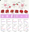
Si-RBCs for liver NMP. (A) Simplified schematic of the liver NMP; (B) native- and (C) 10 mM Si-RBCs perfused liver morphology and pathology sections, (scale bars, 100 μm), respectively; (D) portal vein flow, (E) oxygen pressure, (F) pH trends during liver perfusion; (G) hepatic glucose metabolism levels include lactate level and (H) glucose level; (IM) biochemical analysis during NMP including liver function (I) ALT (U/L), (J) AST (U/L) and (K) ALP (U/L), and biliary function (L) BUN (mg/ dL) and (M) bile production (mL/g).
Fig. 7.
Fig. 7.
Si-RBCs for xenotransfusion. (A) Schematic of Si-RBCs for xenotransfusion; (B) biochemical analysis of liver function, i.e., ALT (U/L), AST (U/L), ALP (U/L), TBIL (U/L); (C) evaluation of Suzuki’s scores for liver injury biochemical analysis of kidney function; (D) pathological sections of liver injury, (scale bars, 200 μm, 100 μm, 50 μm); (E) biochemical analysis of kidney function, i.e., CREA (μM), UREA (mM), UA (μM); (F) pathological sections of kidney injury, (scale bars, 100 μm), (n = 3; mean ± SD).

References

    1. Reddy S. P., Brockmann J., Friend P. J., Normothermic perfusion: A mini-review. Transplantation 87, 631–632 (2009). - PMC - PubMed
    1. Guarrera J. V., et al. , Hypothermic machine preservation in human liver transplantation: The first clinical series. Am. J. Transplant. 10, 372–381 (2010). - PubMed
    1. Thompson E. R., Connelly C., Ali S., Sheerin N. S., Wilson C. H., Cell therapy during machine perfusion. Transpl. Int. 34, 49–58 (2021). - PubMed
    1. Nasralla D., et al. , A randomized trial of normothermic preservation in liver transplantation. Nature 557, 50–56 (2018). - PubMed
    1. Eshmuminov D., et al. , An integrated perfusion machine preserves injured human livers for 1 week. Nat. Biotechnol. 38, 189–198 (2020). - PMC - PubMed

LinkOut - more resources