

The Evolving Portrait of γδ TCR Recognition Determinants
- PMID: 39159405
- PMCID: PMC11335310
- DOI: 10.4049/jimmunol.2400114
The Evolving Portrait of γδ TCR Recognition Determinants
Abstract
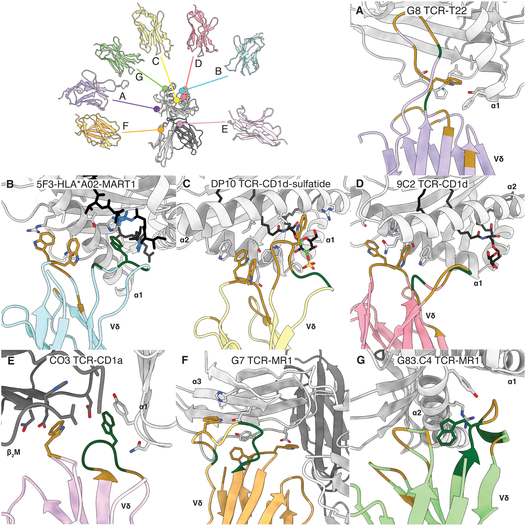
In αβ T cells, immunosurveillance is enabled by the αβ TCR, which corecognizes peptide, lipid, or small-molecule Ags presented by MHC- and MHC class I-like Ag-presenting molecules, respectively. Although αβ TCRs vary in their Ag recognition modes, in general they corecognize the presented Ag and the Ag-presenting molecule and do so in an invariable "end-to-end" manner. Quite distinctly, γδ T cells, by way of their γδ TCR, can recognize ligands that extend beyond the confines of MHC- and MHC class I-like restrictions. From structural studies, it is now becoming apparent that γδ TCR recognition modes can break the corecognition paradigm and deviate markedly from the end-to-end docking mechanisms of αβ TCR counterparts. This brief review highlights the emerging portrait of how γδ TCRs can recognize diverse epitopes of their Ags in a manner reminiscent to how Abs recognize Ags.
Copyright © 2024 by The American Association of Immunologists, Inc.
Conflict of interest statement
Disclosures
The authors declare no competing interests.
Figures

References
-
- Brigl M, and Brenner MB. 2004. CD1: antigen presentation and T cell function. Annu Rev Immunol 22: 817–890. - PubMed
-
- Patel O, Kjer-Nielsen L, Le Nours J, Eckle SB, Birkinshaw R, Beddoe T, Corbett AJ, Liu L, Miles JJ, Meehan B, Reantragoon R, Sandoval-Romero ML, Sullivan LC, Brooks AG, Chen Z, Fairlie DP, McCluskey J, and Rossjohn J. 2013. Recognition of vitamin B metabolites by mucosal-associated invariant T cells. Nat Commun 4: 2142–2151. - PubMed
-
- Hayday AC 2000. [gamma][delta] cells: a right time and a right place for a conserved third way of protection. Annu Rev Immunol 18: 975–1026. - PubMed
-
- Morita CT, Jin C, Sarikonda G, and Wang H. 2007. Nonpeptide antigens, presentation mechanisms, and immunological memory of human Vgamma2Vdelta2 T cells: discriminating friend from foe through the recognition of prenyl pyrophosphate antigens. Immunol Rev 215: 59–76. - PubMed
-
- Costa G, Loizon S Fau - Guenot M, Guenot M Fau - Mocan I, Mocan I Fau - Halary F, Halary F Fau - de Saint-Basile G, de Saint-Basile G Fau - Pitard V, Pitard V Fau - Déchanet-Merville J, Déchanet-Merville J Fau - Moreau J-F, Moreau Jf Fau - Troye-Blomberg M, Troye-Blomberg M Fau - Mercereau-Puijalon O, Mercereau-Puijalon O Fau - Behr C, and Behr C. 2011. Control of Plasmodium falciparum erythrocytic cycle: γδ T cells target the red blood cell-invasive merozoites. Blood 118: 6952–6962. - PubMed
Publication types
MeSH terms
Substances
Grants and funding
LinkOut - more resources
Full Text Sources
Research Materials
