. 2024 Aug 19;8(8):CASE2457.
doi: 10.3171/CASE2457. Print 2024 Aug 19.

Spinal epidural arteriovenous fistula with an intraosseous shunt arising in a compression fracture vertebra: illustrative case

Affiliations

Spinal epidural arteriovenous fistula with an intraosseous shunt arising in a compression fracture vertebra: illustrative case

Natsuki Akaike et al. J Neurosurg Case Lessons. .

Abstract

Background: Spinal epidural arteriovenous fistulas (SEAVFs) with intraosseous shunts are rare, and their underlying pathophysiological mechanisms remain unclear.

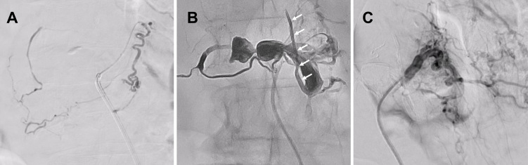
Observations: A female in her 70s presented with rapidly progressive weakness in both lower extremities and urinary retention. Lumbar spine magnetic resonance imaging revealed spinal cord edema and flow voids due to venous dilation and compression fractures of the L1 and L2 vertebral bodies. Spinal angiography revealed ventral and dorsal somatic branches of the lumbar arteries at L1 and L2 flowing into the shunt. High-resolution cone-beam computed tomography revealed a shunt within the compression-fractured vertebral body bone of L2. The intravertebral shunt blood flowed into the ventral epidural venous plexus (VEVP) and returned into the perimedullary vein (PMV). Transarterial embolization was performed using N-butyl cyanoacrylate and Onyx-18 for feeder L1 and feeder L2, respectively. Onyx-18 was injected from the VEVP into the PMV, and complete occlusion of the shunt was achieved. The patient showed symptomatic improvement postoperatively.

Lessons: Vertebral compression fractures are common but rarely associated with SEAVFs. https://thejns.org/doi/10.3171/CASE2457.

Keywords: compression fracture; embolization; spinal epidural arteriovenous fistula.


Figures

FIG. 1.
FIG. 1.
A: Sagittal T2-weighted MRI showing a high signal suggestive of spinal cord congestion and a flow void around the spinal cord (arrows). B: Sagittal T2-weighted MRI showed compression fractures in the L1 and L2 vertebrae, with bony erosion within the L2 vertebra (arrowhead).
FIG. 2.
FIG. 2.
Left L1 arteriography, anteroposterior (A) and lateral (B) projections, showing the SEAVF (white arrow) fed by the ventral and dorsal somatic branches of the lumbar artery and a venous pouch, with shunt blood flow into the VEVP and back into the PMV (black arrow). Left L2 lumbar arteriography, anteroposterior projection (C), showing a high-flow SEAVF (white arrow) fed by the ventral and dorsal somatic branches of the lumbar artery and a venous pouch, with shunt blood flow into the VEVP and back into the PMV (black arrow). Axial high-resolution cone-beam CT image showing a compression fracture of the L2 vertebra, with internal bone erosion (white asterisk, D). Dorsal and ventral somatic branches are depositing blood into a shunt, and a venous pouch (black asterisk, E) is consistent with the bone erosion image.
FIG. 3.
FIG. 3.
A: TAE was performed using 20% NBCA for the feeder from the lumbar artery. B: The Rebar catheter was guided posteriorly to the vertebral body near the shunt through the L2 segmental artery, from which Onyx-18 was injected. TAE was performed using Onyx-18 and injected from the VEVP to the PMV (white arrows). C: The final angiogram revealed complete obliteration of the fistula.
FIG. 4.
FIG. 4.
Postoperative lumbar MRI showing the disappearance of the flow void.

References

    1. Kiyosue H, Matsumaru Y, Niimi Y, et al. Angiographic and clinical characteristics of thoracolumbar spinal epidural and dural arteriovenous fistulas. Stroke. 2017;48(12):3215-3222. - PMC - PubMed
    1. Brinjikji W, Yin R, Nasr DM, Lanzino G. Spinal epidural arteriovenous fistulas. J Neurointerv Surg. 2016;8(12):1305-1310. - PubMed
    1. Kiyosue H, Tanoue S, Okahara M, Hori Y, Kashiwagi J, Mori H. Spinal ventral epidural arteriovenous fistulas of the lumbar spine: angioarchitecture and endovascular treatment. Neuroradiology. 2013;55(3):327-336. - PMC - PubMed
    1. Chul Suh D, Gon Choi C, Bo Sung K, Kim KK, Chul Rhim S. Spinal osseous epidural arteriovenous fistula with multiple small arterial feeders converging to a round fistular nidus as a target of venous approach. AJNR Am J Neuroradiol. 2004;25(1):69-73. - PMC - PubMed
    1. Gjertsen O, Nakstad PH, Pedersen H, Dahlberg D. Percutaneous intravertebral body embolization of a traumatic spinal epidural arteriovenous fistula with secondary perimedullary venous reflux. A case report. Interv Neuroradiol. 2010;16(1):97-102. - PMC - PubMed