. 2024 Aug 19;15(1):7113.
doi: 10.1038/s41467-024-51169-0.

A lactobacilli-based inhaled live biotherapeutic product attenuates pulmonary neutrophilic inflammation

Affiliations

A lactobacilli-based inhaled live biotherapeutic product attenuates pulmonary neutrophilic inflammation

Teodora Nicola et al. Nat Commun. .

Abstract

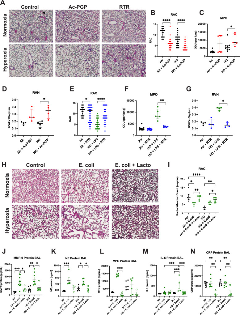
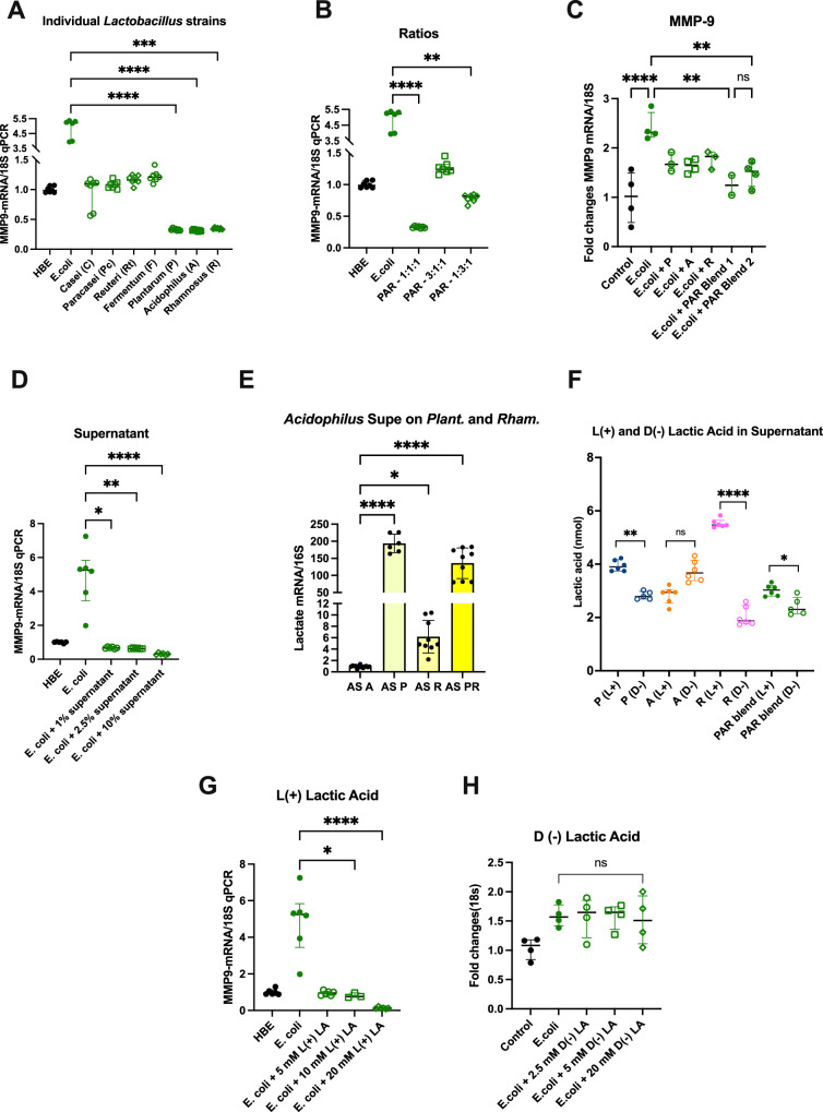
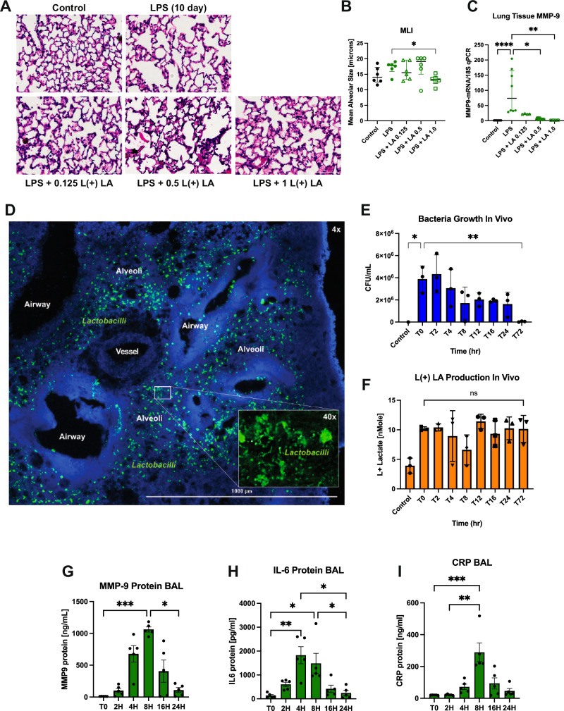
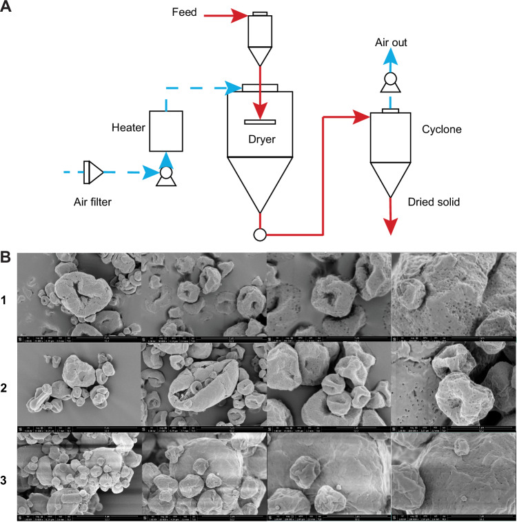
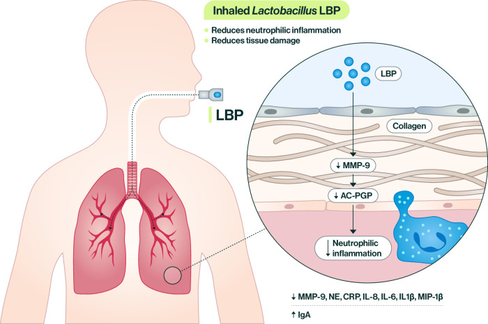
Bronchopulmonary dysplasia (BPD) is a chronic lung disease of prematurity. Exposure to noxious stimuli such as hyperoxia, volutrauma, and infection in infancy can have long-reaching impacts on lung health and predispose towards the development of conditions such as chronic obstructive pulmonary disease (COPD) in adulthood. BPD and COPD are both marked by lung tissue degradation, neutrophil influx, and decreased lung function. Both diseases also express a change in microbial signature characterized by firmicute depletion. However, the relationship between pulmonary bacteria and the mechanisms of downstream disease development has yet to be elucidated. We hypothesized that murine models of BPD would show heightened acetylated proline-glycine-proline (Ac-PGP) pathway and neutrophil activity, and through gain- and loss-of-function studies we show that Ac-PGP plays a critical role in driving BPD development. We further test a inhaled live biotherapeutic (LBP) using active Lactobacillus strains in in vitro and in vivo models of BPD and COPD. The Lactobacillus-based LBP is effective in improving lung structure and function, mitigating neutrophil influx, and reducing a broad swath of pro-inflammatory markers in these models of chronic pulmonary disease via the MMP-9/PGP (matrix metalloproteinase/proline-glycine-proline) pathway. Inhaled LBPs show promise in addressing common pathways of disease progression that in the future can be targeted in a variety of chronic lung diseases.


Conflict of interest statement

Part of the research described in this manuscript is patented under “Inhaled Respiratory Probiotics for Lung Diseases of Infancy, Childhood and Adulthood” US 11,141,443 B2 held under the University of Alabama at Birmingham Research Foundation (CL, AG, NA are inventors). The remaining authors declare no competing interests.

Figures

Fig. 1
Fig. 1. Severe BPD is marked by decreased Lactobacilli, increased proteobacteria, and neutrophil influx mediated by the MMP-9/Ac-PGP pathway.
A Infants with severe BPD had increased proteobacteria, decreased firmicutes, and increased endotoxin levels compared to controls as measured by 16s microbiome sequencing (P = 0.0284) (data from ref. ). Infants with severe BPD had higher concentrations of B Ac-PGP (P = 0.0003), C MMP-9 (P = 0.0002), D MPO (P = 0.0023; Control N = 10, BPD N = 7 samples), and E NE protein (P < 0.0001; Control N = 16, BPD N = 8 samples) in their tracheal aspirates than controls. Unpaired t-test. Ac-PGP was measured by tandem mass spectrometry; MMP-9, MPO, and NE were measured by ELISA. Control N = 7, BPD N = 7 samples. Mice were exposed to hyperoxia (HO) from PN3- PN14, LPS on PN3, 6, 9, and 12, and euthanized on PN14. F Mice exposed to HO + LPS demonstrated severe alveolar hypoplasia and simplification. Tissue slices from harvested lung tissue were photographed at 4x magnification after hematoxylin and eosin (H&E) staining. G Radial alveolar count (RAC) decreased in LPS mice. N = 24 samples. Pulmonary function worsened as measured by H resistance (increased) (Air N = 7, Air+LPS N = 8, HO N = 5, HO + LPS N = 7 mice) and I compliance (decreased) in HO + LPS mice (Air N = 10, Air+LPS N = 8, HO N = 5, HO + LPS N = 7 mice). LPS-exposed mice had higher concentrations of J Ac-PGP (Air N = 8, other groups N = 7 mice) and K MPO in the bronchoalveolar lavage fluid (BAL) (Air N = 10, Air + LPS N = 7, HO N = 6, HO + LPS N = 5 mice). One-way ANOVA, Tukey’s multiple comparisons test. Bars represent the median ± interquartile range. *P < 0.05, **P < 0.01, ***P < 0.001, ****P < 0.0001.
Fig. 2
Fig. 2. Ac-PGP drives tissue damage and Lacto LBP reduces neutrophilic inflammation.
Ac-PGP and LPS exposure each in combination with hyperoxia (HO) resulted in severe alveolar hypoplasia and simplification. A Treatment with RTR (arginine-threonine-arginine) improved alveolar structure. H&E staining, 4x magnification. B RAC decreased upon dosing with Ac-PGP or LPS (N = 120 samples) and C MPO expression increased in Ac-PGP exposure in HO (P = 0.0451; Air N = 11, Air+Ac-PGP N = 8, HO N = 8, HO+Ac-PGP N = 5 mice). D Right ventricular hypertrophy (RVH) increased in Ac-PGP + HO mice. (P = 0.028; N = 4 mice). E RAC improved upon treatment with RTR. N = 120 samples. F MPO expression increased upon exposure to LPS + HO and reduced upon RTR treatment (P = 0.0079; Air N = 10, Air + RTR N = 9, HO N = 5, HO + LPS + RTR N = 5 mice). G RVH decreased in LPS + HO mice treated with RTR (P = 0.0187; N = 4 mice). Mann–Whitney U-test, Kruskal–Wallis, Dunn’s multiple comparisons test. H Hyperoxia exacerbated alveolar simplification in mice exposed to E. coli intranasally, while intratracheal LBP treatment improved tissue structure in E. coli + HO mice. H&E staining, 4x magnification. I RAC decreased in HO + E. coli exposure and restored to normal with LBP treatment. Air N = 7, Air + E. coli N = 5, Air + E. coli + Lacto N = 5, HO groups N = 5 mice each. J BAL MMP-9, K NE, L MPO, M IL-6, and N CRP decreased upon treatment with LBP. One-way ANOVA, Tukey’s multiple comparisons test. Air N = 7, Air + E. coli N = 10, Air + E. coli + Lacto N = 5, HO N = 6, HO + E. coli N = 8, HO + E. coli + Lacto N = 5 mice. Bars represent the median ± interquartile range. *P < 0.05, **P < 0.01, ***P < 0.001, ****P < 0.0001.
Fig. 3
Fig. 3. Lactobacillus blend reduces neutrophilic inflammation through L (+) lactic acid production.
A Individual strains L. plantarum (P), L. acidophilus (A), and L. rhamnosus (R) reduce MMP-9 expression more than individual strains L. casei, L. paracasei, L. reuteri, and L. fermentum compared to E. coli exposure alone with no Lactobacillus treatment and compared to each other. N = 7. B Different ratios of blended P, A, and R reduce MMP-9 to varying degrees. N = 7 wells. C A blend of P, A, and R performed better than individual strains in reducing MMP-9 in human bronchial epithelial (HBE) in vitro model of noxious stimuli exposure. N = 4 wells. D Lactobacillus blend culture supernatant in increasing concentrations reduces MMP-9 expression. N = 6. E Individual live Lactobacillus strains were transfected with supernatant from the other three strains in the blend. L. acidophilus supernatant cultured with live A, P, R, and PR blend increased lactate gene expression of P and the PR blend (shown as a representative example). AS A N = 13, AS P N = 6, AS R N = 9, AS PR N = 9 wells. F Lactobacillus strains produce more L (+) lactic acid than D (+) lactic acid individually and as a blend. N = 6 wells. G L (+) lactic acid reduces MMP-9 expression in HBE cells exposed to E. coli (N = 6 wells), while H D (−) lactic acid does not (N = 4 wells). Kruskal–Wallis test, Dunn’s multiple comparisons. Cell culture performed in triplicate. Bars represent the median ± interquartile range. *P < 0.05, **P < 0.01, ***P < 0.001, ****P < 0.0001.
Fig. 4
Fig. 4. L (+) lactic acid reduces MMP-9 and is produced by Lactobacilli in vivo.
Mice exposed to LPS intratracheally for 10 days were dosed with L (+) lactic acid in the lungs. A Lung histology images show alveolar simplification upon LPS injury and slight recovery upon LA treatment. H&E staining, 4x magnification. B MLI decreased upon treatment with 1 ug/g body weight L (+) LA per mouse (P = 0.0152; N = 6 mice). C Lung tissue MMP-9 expression decreased upon IT L (+) lactic acid treatment (Control N = 10, LPS N = 8, LA 0.125 N = 4, LA 0.5 N = 12, LA 1.0 N = 4 mice). D Lactobacilli (green) localizes to the lung epithelium 3 h after intratracheal inoculation to healthy mice. DAPI blue. 4x magnification, 40x magnification on inset. N = 5. E Healthy mice inoculated with Lactobacillus LBP to the lungs show progressive clearance of bacteria via the reduction in colonies over 72 h. F Healthy mice inoculated with LBP to the lungs show an increase in L (+) lactic acid production and sustained effect through 72 h post-dose. L(+) LA measured by lactate colorimetric assay. N = 3 mice at each time point. GI Inflammatory biomarkers MMP-9, CRP, and IL-6 were elevated at 4–8 h post-LBP dose and returned to baseline levels by 16–24 h. N = 5 mice at each time point. Mann–Whitney U-test and Kruskal–Wallis, Dunn’s multiple comparisons. Bars represent the median ± interquartile range. *P < 0.05, **P < 0.01, ***P < 0.001, ****P < 0.0001.
Fig. 5
Fig. 5. Spray drying produces small, flowable powder particles containing viable bacteria.
A Schematic of spray drying process used to dry Lactobacillus strains into small, flowable powder particles. B Scanning electron microscope (SEM) images of three formulations of powder particles after drying. Images at 25,000, 50,000, and 100,000X magnification (left to right) were taken from one sample each.
Fig. 6
Fig. 6. Lactobacillus-based LBP improves biomarkers of inflammation and lung structure and function in murine models of COPD.
Mice were exposed to cigarette smoke for 1 month and treated intratracheally with the Lactobacillus LBP (Lacto). A Lung histology image (H&E staining, 40x magnification) did not show significant worsening of alveolar structure upon 1 month of smoke exposure as measured by (B) and mean linear intercept (MLI). Measurements: Air N = 5, Air + Lacto N = 11, Smoke N = 23, Smoke + Lacto N = 27 samples. Mice exposed to smoke treated with Lacto showed improvements in (C) lung tissue MMP-9 expression (Air N = 4, Smoke N = 5 mice), D serum MMP-9 protein levels (Air N = 4, Smoke N = 5 mice), and E BAL IgA protein levels (Air N = 4, Smoke N = 6 mice). Mice were exposed to intratracheal porcine pancreatic elastase (PPE) and LPS, leading to significant alveolar hypoplasia and simplification. F Lung histology images (H&E staining, 4x magnification) showed that inhalation of the Lacto LBP concurrent with the injury period reduced tissue damage. G MLI improved upon treatment with Lacto LBP in PPE and PPE + LPS exposure groups. H Lung function worsened as measured by increased resistance upon exposure to PPE and improved upon treatment with Lacto LBP. I Lung function, as measured by compliance, showed no significant changes among groups. J MMP-9 expression in lung tissue increased upon PPE + LPS exposure and decreased upon treatment with Lacto LBP. K MMP-9 protein in the BAL increased upon PPE and PPE + LPS exposure and decreased upon treatment with Lacto LBP in PPE + LPS mice. L NE protein in the BAL decreased upon Lacto LBP treatment in PPE + LPS mice. M CRP protein in the BAL increased upon PPE + LPS exposure and decreased upon treatment with Lacto LBP. N IL-8 protein in the BAL decreased upon Lacto LBP treatment in PPE and PPE + LPS mice. O IgA protein in the BAL increased (improved) upon Lacto LBP treatment in control, PPE, and PPE + LPS mice. N = 12 mice (6 M/6 F). Kruskal–Wallis test, Dunn’s multiple comparisons. Bars represent the median ± interquartile range. *P < 0.05, **P < 0.01, ***P < 0.001, ****P < 0.0001.
Fig. 7
Fig. 7. Mice were exposed to intratracheal PPE and LPS to establish emphysema and bacteria-driven inflammation.
A Lung histology images (H&E staining, 4x magnification) show that Lacto LBP treatment after 2 weeks of injury was established, reduced inflammation, and improved tissue structure. Steroid treatment after injury decreases inflammation. B PPE and PPE + LPS exposure each increased mean alveolar size and Lacto LBP treatment significantly decreased (improved) it. The steroid fluticasone furoate (FF) also significantly reduced MLI but not as much as Lacto LBP in the PPE group. Measurements: Excipient N = 16, Lacto N = 8, Steroid N = 18, PPE + Excipient N = 44, PPE + Lacto N = 18, PPE + Steroid N = 18, PPE + LPS N = 13, PPE + LPS + Lacto N = 24, PPE + LPS + Steroid N = 23 samples. C Lacto LBP performed better than the steroid in reducing MMP-9 protein in BAL of mice exposed to PPE + LPS. D Lacto LBP performed as well as the steroid in reducing CRP protein in BAL of mice exposed to PPE and PPE + LPS. Excipient N = 5, Lacto N = 2, Steroid N = 3, PPE + Excipient N = 3, PPE + Lacto N = 6, PPE + Steroid N = 4, PPE + LPS N = 5, PPE + LPS + Lacto N = 7, PPE + LPS + Steroid N = 6 mice. Kruskal–Wallis test, Dunn’s multiple comparisons. Bars represent the median ± interquartile range. *P < 0.05, **P < 0.01, ***P < 0.001, ****P < 0.0001.
Fig. 8
Fig. 8. Inhaled Lactobacillus-based LBP shows favorable safety and biodistribution profile in the PPE mouse model.
A 100% of the mice in the biodistribution study survived at 23 days (terminal sacrifice) and 28 days (recovery sacrifice). Survival curves offset for visibility. Body weight, temperature, oxygen saturation, breath rate, breath distension, heart rate, and pulse distension remained in normal ranges after 2 weeks of LBP dosing in BH terminal sacrifice mice and IO recovery sacrifice mice. P Representative gels from PCR on terminal sacrifice tissues lung, serum, and brain show no presence of LBP Lactobacillus strains. Q Representative gels from PCR on recovery sacrifice tissues lung, serum, and brain show no presence of LBP Lactobacillus strains. PBS and PPE control each N = 10; PPE + placebo, LBP 1 mg, LBP 3 mg each N = 15 mice. Kruskal–Wallis test, Dunn’s multiple comparisons. Bars represent the median ± interquartile range. *P < 0.05, **P < 0.01, ***P < 0.001, ****P < 0.0001.
Fig. 9
Fig. 9. Schematic diagram of how the inhaled Lactobacillus LBP works.
Particle engineering innovation facilitates the delivery of the Lactobacillus LBP directly into the lung to help reduce inflammation and restore lung tissue. Inhaled LBP releases anti-inflammatory metabolites that reduce MMP-9 and Ac-PGP expression and neutrophilic inflammation downstream. Other inflammatory biomarkers are reduced, and protective IgA is increased.

References

    1. Kalikkot Thekkeveedu, R., Guaman, M. C. & Shivanna, B. Bronchopulmonary dysplasia: a review of pathogenesis and pathophysiology. Respir. Med.132, 170–177 (2017). 10.1016/j.rmed.2017.10.014 - DOI - PMC - PubMed
    1. Lohmann, P. et al. The airway microbiome of intubated premature infants: characteristics and changes that predict the development of bronchopulmonary dysplasia. Pediatr. Res.76, 294–301 (2014). 10.1038/pr.2014.85 - DOI - PubMed
    1. Bhandari, A. & Bhandari, V. Pathogenesis, pathology and pathophysiology of pulmonary sequelae of bronchopulmonary dysplasia in premature infants. Front. Biosci.8, e370–e380 (2003). 10.2741/1060 - DOI - PubMed
    1. Glass, H. C. et al. Outcomes for extremely premature infants. Anesth. Analg.120, 1337–1351 (2015). 10.1213/ANE.0000000000000705 - DOI - PMC - PubMed
    1. Stocks, J. & Sonnappa, S. Early life influences on the development of chronic obstructive pulmonary disease. Ther. Adv. Respir. Dis.7, 161–173 (2013). 10.1177/1753465813479428 - DOI - PMC - PubMed

MeSH terms