. 2024 Aug 20;14(1):19225.
doi: 10.1038/s41598-024-69696-7.

Human glioblastoma-derived cell membrane nanovesicles: a novel, cell-specific strategy for boron neutron capture therapy of brain tumors

Affiliations

Human glioblastoma-derived cell membrane nanovesicles: a novel, cell-specific strategy for boron neutron capture therapy of brain tumors

Alice Balboni et al. Sci Rep. .

Abstract

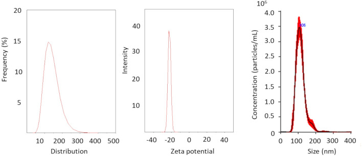
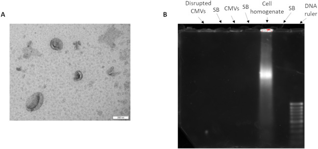
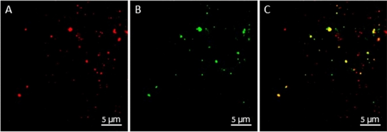
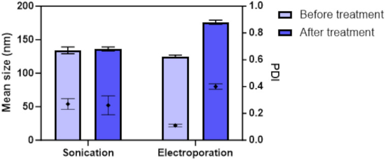
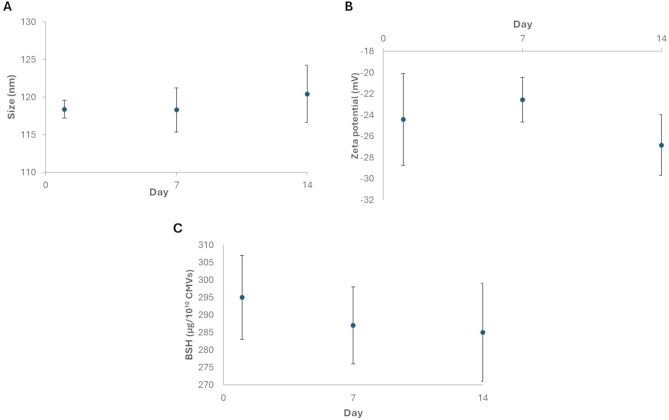
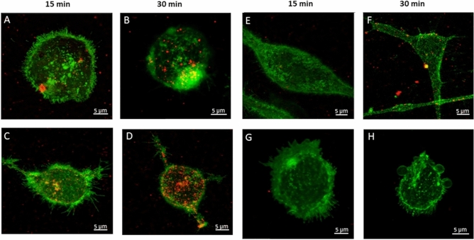
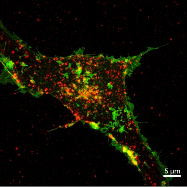
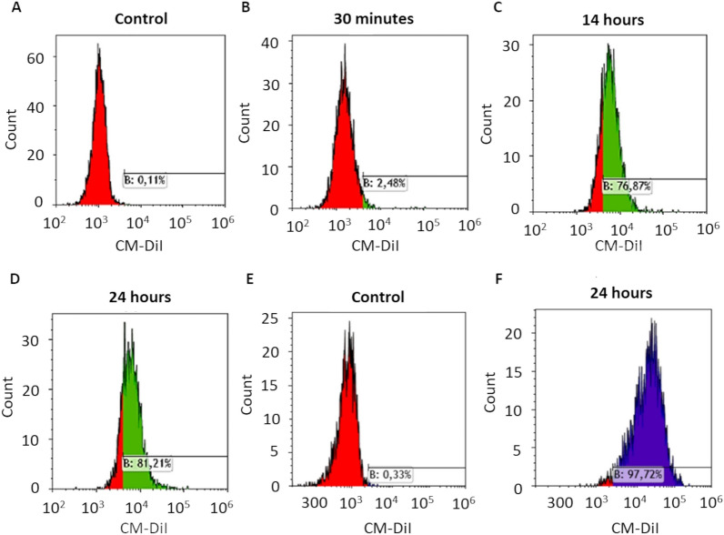
Glioblastoma (GBM), one of the deadliest brain tumors, accounts for approximately 50% of all primary malignant CNS tumors, therefore novel, highly effective remedies are urgently needed. Boron neutron capture therapy, which has recently repositioned as a promising strategy to treat high-grade gliomas, requires a conspicuous accumulation of boron atoms in the cancer cells. With the aim of selectively deliver sodium borocaptate (BSH, a 12 B atoms-including molecule already employed in the clinics) to GBM cells, we developed novel cell membrane-derived vesicles (CMVs), overcoming the limits of natural extracellular vesicles as drug carriers, while maintaining their inherent homing abilities that make them preferable to fully synthetic nanocarriers. Purified cell membrane fragments, isolated from patient-derived GBM stem-like cell cultures, were used to prepare nanosized CMVs, which retained some membrane proteins specific of the GBM parent cells and were devoid of potentially detrimental genetic material. In vitro tests evidenced the targeting ability of this novel nanosystem and ruled out any cytotoxicity. The CMVs were successfully loaded with BSH, by following two different procedures, i.e. sonication and electroporation, demonstrating their potential applicability in GBM therapy.

Keywords: Bioinspired vesicles; Cancer treatment; Cell internalization; Cell membrane; Proteomics.


Conflict of interest statement

The authors declare that they have no known competing financial interests or personal relationships that could have appeared to influence the work reported in this paper.

Figures

Figure 1
Figure 1
On the left: CMV size distribution by intensity; in the middle: CMV zeta potential; on the right: CMV concentration vs size distribution measured by NTA (sample dilution 1:500).
Figure 2
Figure 2
(A) TEM micrograph of the CMVs, magnification bar = 200 nm; (B) Agarose gel electrophoresis revealing the absence of genetic material in CMVs, even when disrupted by ultrasonication. A slight overexposure was applied to the gel to further validate the absence of genetic material in the CMV samples.
Figure 3
Figure 3
Confocal images of CM-DiI-labeled CMVs after incubation with anti-annexin A2 antibody labeled with the Alexa Fluor 488 dye. (A) Red fluorescent signal deriving from CM-DiI-labelled CMVs (λex = 561 nm); (B) Green fluorescent signal deriving from the Alexa Fluor 488-labelled anti-annexin A2 antibody (λex = 488 nm); (C) Merged image, highlighting the co-localization of the two dyes.
Figure 4
Figure 4
Mean CMV size and PDI values before and after BSH loading, carried out by sonication and electroporation. Mean ± standard deviation (n = 3).
Figure 5
Figure 5
Stability of the BSH-loaded CMVs over 2 week storage at 4 °C, monitoring the size (A), zeta potential (B) and BSH content (C).
Figure 6
Figure 6
Confocal images obtained by the superimposition of different cell stacks (0.7 µm height). CM-DiI-labeled CMVs (red) incubated for 15 and 30 min with the green fluorescent parent GBM1 cells (A and B), non-parent GBM2 cells (C and D), and human M03.13 oligodendrocytes (E and F). CM-DiI-labeled liposomes were also incubated with GBM1 cells (G and H), to rule out a non-specific interaction of phospholipids with GBM cells. Scale bar 5 µm.
Figure 7
Figure 7
Confocal image obtained by the superimposition of different cell stacks (0.7 µm height). CM-DiI-labeled and BSH-loaded CMVs (red) incubated for 30 min with the green fluorescent parent GBM1 cells.
Figure 8
Figure 8
Flow cytometry histograms representing relative CM-DiI fluorescence intensity (x-axis) and the number of events (count, y-axis). Parent GBM1 cells not incubated with CM-DiI labeled CMVs (A), incubated with CM-DiI-labeled CMVs for 30 min (B), with CM-DiI-labeled CMVs for 16 h (C) and with CM-DiI-labeled CMVs for 24 h (D); non-parent GBM2 cells not incubated with CM-DiI labeled CMVs (E), and incubated with CM-DiI-labeled CMVs for 24 h (F).
Figure 9
Figure 9
Cell viability in control and CMV-treated cells assessed by MTT assay on human M03.13 oligodendrocyte cell line (A), parent GBM1 cells (B) and non-parent GBM2 cells (C). Two different CMVs concentrations and two time points were tested. *p < 0.05; **p < 0.01; ***p < 0.001 versus control.

References

    1. Maher, E. A. et al. Malignant glioma: Genetics and biology of a grave matter. Genes Dev.15, 1311–1333 (2001). 10.1101/gad.891601 - DOI - PubMed
    1. Florio, T. & Barbieri, F. The status of the art of human malignant glioma management: The promising role of targeting tumor-initiating cells. Drug Discov. Today17, 1103–1110 (2012). 10.1016/j.drudis.2012.06.001 - DOI - PubMed
    1. Ostrom, Q. T. et al. The epidemiology of glioma in adults: A ‘state of the science’ review. Neuro-Oncology16, 896–913 (2014). 10.1093/neuonc/nou087 - DOI - PMC - PubMed
    1. Grochans, S. et al. Epidemiology of glioblastoma multiforme-literature review. Cancers14, 2412 (2022). 10.3390/cancers14102412 - DOI - PMC - PubMed
    1. Furnari, F. B. et al. Malignant astrocytic glioma: Genetics, biology, and paths to treatment. Genes Dev.21, 2683–2710 (2007). 10.1101/gad.1596707 - DOI - PubMed

LinkOut - more resources