. 2024 Aug 5:12:1428988.
doi: 10.3389/fbioe.2024.1428988. eCollection 2024.

Antibacterial and antioxidant phlorizin-loaded nanofiber film effectively promotes the healing of burn wounds

Affiliations

Antibacterial and antioxidant phlorizin-loaded nanofiber film effectively promotes the healing of burn wounds

Ying Yang et al. Front Bioeng Biotechnol. .

Abstract

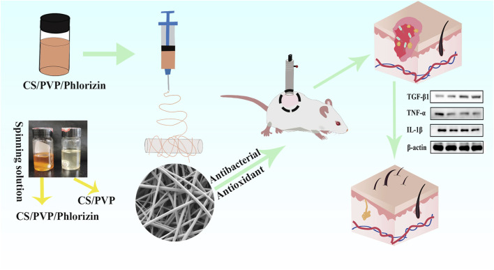
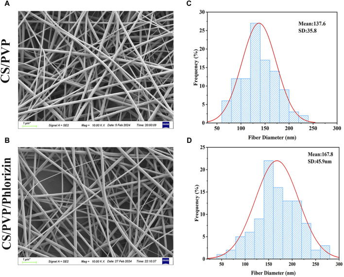
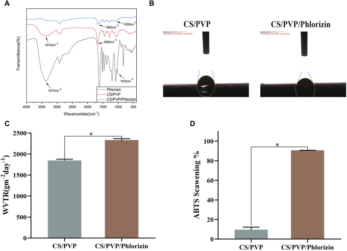
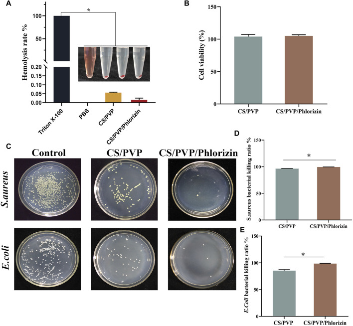
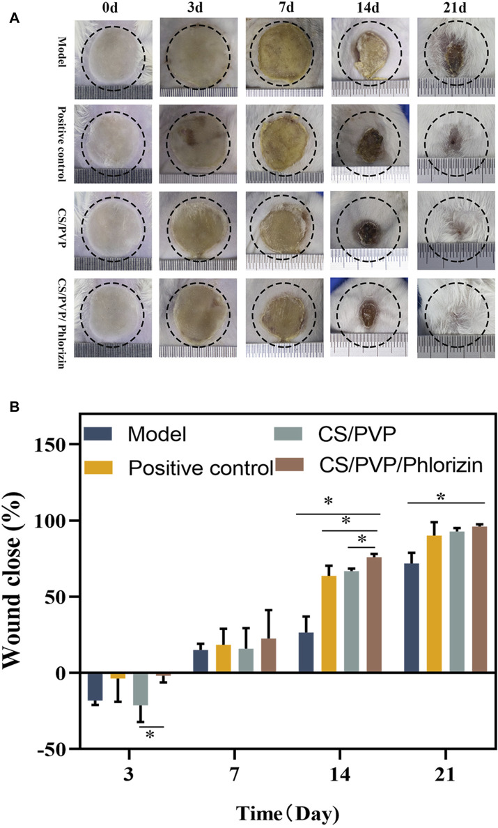
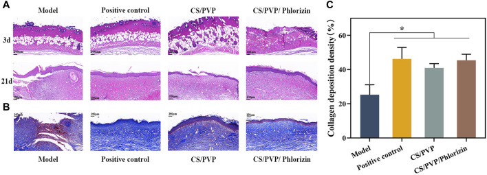
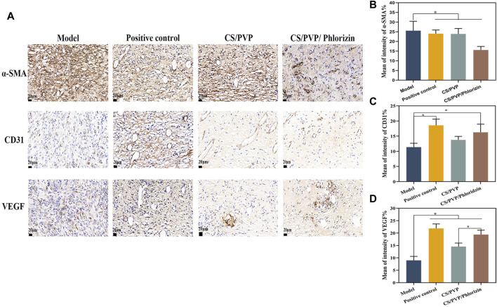
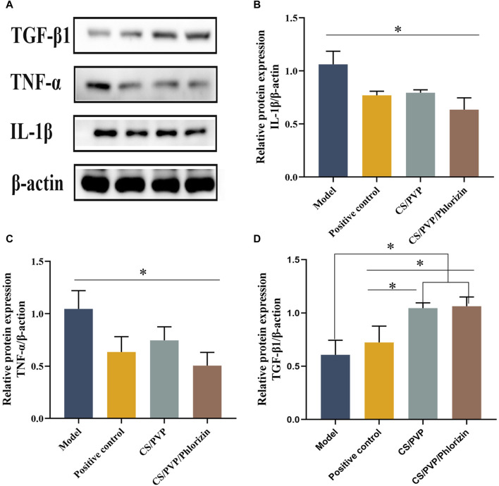
Burns usually result in damage and loss of skin forming irregular wound wounds. The lack of skin tissue protection makes the wound site highly vulnerable to bacterial infections, hindering the healing process. However, commonly used wound dressings do not readily provide complete coverage of irregular wounds compared to regular wounds. Therefore, there is an urgent need to prepare a wound dressing with high antimicrobial efficacy for the administration of drugs to irregular wounds. In this study, a chitosan (CS)/polyvinylpyrrolidone (PVP) composite nanofiber membrane (CS/PVP/Phlorizin) loaded with root bark glycosides (Phlorizin) was developed using an electrostatic spinning technique. The incorporation of phlorizin, a natural antioxidant, into the fiber membranes notably boosted their antimicrobial and antioxidant capabilities, along with demonstrating excellent hydrophilic characteristics. In vitro cellular experiments showed that CS/PVP/Phlorizin increased Hacat cell viability with the presence of better cytocompatibility. In scald wound healing experiments, Phlorizin-loaded nanofibrous membranes significantly promoted re-epithelialization and angiogenesis at the wound site, and reduced the inflammatory response at the wound site. Therefore, the above results indicate that this nanofiber membrane is expected to be an ideal dressing for burn wounds.

Keywords: chitosan; nanofiber membrane; phlorizin; scalding wound repair; wound dressing.


Conflict of interest statement

Authors AL, GX, CD, XS, LY, and MY were employed by Jilin Aodong Yanbian Pharmaceutical Co, Ltd. The remaining authors declare that the research was conducted in the absence of any commercial or financial relationships that could be construed as a potential conflict of interest.

Figures

SCHEME 1
SCHEME 1
Loaded phlorizin nanofiber membrane to promote scald wound healing
FIGURE 1
FIGURE 1
SEM image of nanofiber membrane and its nanofiber diameter size. (A, B) SEM images of CS/PVP, CS/PVP/Phlorizin nanofiber membranes; (C, D) Diameter distribution of CS/PVP, CS/PVP/Phlorizin nanofiber membranes, results were used as mean ± standard deviation.
FIGURE 2
FIGURE 2
(A) Fourier infrared spectra; (B) water contact angle; (C) water vapor transmission rate; (D) ABTS radical scavenging rate.
FIGURE 3
FIGURE 3
(A) Hemocompatibility; (B) Cytotoxicity; (C) Colony counts of CS/PVP, CS/PVP/Phlorizin nanofiber membranes for Staphylococcus aureus and Escherichia coli; (D) Quantitative graph of the inhibition rate of the nanofiber membranes for Staphylococcus aureus; (E) Quantitative graph of the inhibition rate of the nanofiber membranes for Escherichia coli.
FIGURE 4
FIGURE 4
(A) Wound status and its size change on days 0, 3, 7, 4, and 21 for Model, Positive control, CS/PVP, and CS/PVP/Phlorizin groups; (B) Quantitative plot of wound close rate.
FIGURE 5
FIGURE 5
(A) H&E staining of Model, Positive control, CS/PVP, and CS/PVP/Phlorizin groups on days 3 and 21; (B) Masson staining of each group on day 21; (C) collagen deposition statistics of each group.
FIGURE 6
FIGURE 6
(A) Immunohistochemical pictures of α-SMA, CD31, and VEGF in each group; (B) Quantitative analysis pictures of α-SMA; (C) Quantitative analysis pictures of CD31; (D) Quantitative analysis pictures of VEGF.
FIGURE 7
FIGURE 7
(A) Pictures of TGF-β1, TNF-α, and IL-1β protein bands; (B) Pictures of quantified gray values of IL-1β protein; (C) Pictures of quantified gray values of TNF-α protein; (D) Pictures of quantified gray values of TGF-β1 protein.

References

    1. Archana D., Dutta J., Dutta P. K. (2013). Evaluation of chitosan nano dressing for wound healing: characterization, in vitro and in vivo studies. Int. J. Biol. Macromol. 57, 193–203. 10.1016/j.ijbiomac.2013.03.002 - DOI - PubMed
    1. Asiri A., Saidin S., Sani M. H., Al-Ashwal R. H. (2021). Epidermal and fibroblast growth factors incorporated polyvinyl alcohol electrospun nanofibers as biological dressing scaffold. Sci. Rep. 11 (1), 5634. - PMC - PubMed
    1. Boateng J. S., Matthews K. H., Stevens H. N. E., Eccleston G. M. (2008). Wound healing dressings and drug delivery systems: a review. J. Pharm. Sci. 97 (8), 2892–2923. 10.1002/jps.21210 - DOI - PubMed
    1. Contardi M., Kossyvaki D., Picone P., Summa M., Guo X., Heredia-Guerrero J. A., et al. (2021). Electrospun polyvinylpyrrolidone (PVP) hydrogels containing hydroxycinnamic acid derivatives as potential wound dressings. Chem. Eng. J. 409, 128144. 10.1016/j.cej.2020.128144 - DOI
    1. Ding C., Yang J., Wang N., Ding Q., Sun S., Gao Y., et al. (2024). Sodium alginate/polyvinyl alcohol nanofibers loaded with Shikonin for diabetic wound healing: in vivo and in vitro evaluation. Int. J. Biol. Macromol. 262, 129937. 10.1016/j.ijbiomac.2024.129937 - DOI - PubMed

LinkOut - more resources