. 2024 Jul 19:31:100612.
doi: 10.1016/j.phro.2024.100612. eCollection 2024 Jul.

3D Unsupervised deep learning method for magnetic resonance imaging-to-computed tomography synthesis in prostate radiotherapy

Affiliations

3D Unsupervised deep learning method for magnetic resonance imaging-to-computed tomography synthesis in prostate radiotherapy

Blanche Texier et al. Phys Imaging Radiat Oncol. .

Abstract

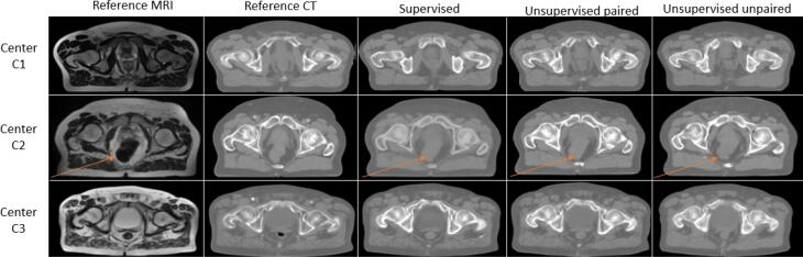
Background and purpose: Magnetic resonance imaging (MRI)-to-computed tomography (CT) synthesis is essential in MRI-only radiotherapy workflows, particularly through deep learning techniques known for their accuracy. However, current supervised methods are limited to specific center's learnings and depend on registration precision. The aim of this study was to evaluate the accuracy of unsupervised and supervised approaches in the context of prostate MRI-to-CT generation for radiotherapy dose calculation.

Methods: CT/MRI image pairs from 99 prostate cancer patients across three different centers were used. A comparison between supervised and unsupervised conditional Generative Adversarial Networks (cGAN) was conducted. Unsupervised training incorporates a style transfer method with. Content and Style Representation for Enhanced Perceptual synthesis (CREPs) loss. For dose evaluation, the photon prescription dose was 60 Gy delivered in volumetric modulated arc therapy (VMAT). Imaging endpoint for sCT evaluation was Mean Absolute Error (MAE). Dosimetric endpoints included absolute dose differences and gamma analysis between CT and sCT dose calculations.

Results: The unsupervised paired network exhibited the highest accuracy for the body with a MAE at 33.6 HU, the highest MAE was 45.5 HU obtained with unsupervised unpaired learning. All architectures provided clinically acceptable results for dose calculation with gamma pass rates above 94 % (1 % 1 mm 10 %).

Conclusions: This study shows that multicenter data can produce accurate sCTs via unsupervised learning, eliminating CT-MRI registration. The sCTs not only matched HU values but also enabled precise dose calculations, suggesting their potential for wider use in MRI-only radiotherapy workflows.

Keywords: Perceptual loss; Synthetic CT; Unsupervised learning; cGAN.


Conflict of interest statement

The authors declare the following financial interests/personal relationships which may be considered as potential competing interests: Cédric Hémon benefits from a PhD scholarship granted by Elekta AB.

Figures

Fig. 1
Fig. 1
Workflow of the study. The workflow of synthetic computed tomography (sCT) generation is as follows: first, images were preprocessed; then, MRIs and CTs are non-rigidly registered for the supervised study. Afterwards, datasets are used to train three cGAN-based networks: a supervised one, an unsupervised unpaired one, and an unsupervised paired one. These networks use different computations of the perceptual loss (PL). Finally, sCTs are evaluated using Mean Absolute Error (MAE), Mean Error (ME), and Peak Signal-to-Noise Ratio (PSNR), and dose evaluation with gamma analysis and Dose Volume Histogram (DVH) comparison is performed.
Fig. 2
Fig. 2
Representation of pelvis CT content extracted by inverse optimization. Similar to Mahendran et al. , an optimization to find an image y^ that minimizes the content reconstruction loss lcontϕ,j(.y^,y) is used.
Fig. 3
Fig. 3
Image results with 3D cGAN according to the chosen learning process: supervised, unsupervised with paired data and unsupervised with unpaired data. The orange arrow shows the presence of gas in the rectum for MRI and the abscence of gas in the sCTs.
Fig. 4
Fig. 4
MAE results on external body: monocentric and multicentric supervised training comparison across centers. The boxplots illustrate the mean absolute error (MAE) between sCT and CT results for when the learning was performed on a cohort: C1 (blue), C2 (yellow), and C3 (red) and the three centers (green) and the test was performed on C1, C2 or C3.
Fig. 5
Fig. 5
sCT dose evaluation results. Section 1 presents the results of the gamma pass rate (GPR) according to the method and the test center. Sections 2, 3 and 4 presents the DVH comparison (respectively absolute differences of mean dose in the bladder, absolute differences of mean dose in the rectum and absolute differences of D95 % in the prostate) results according to the methods (supervised in blue, unsupervised unpaired, in yellow, unsupervised paired, in pink) and the test center.

References

    1. Gao Z., Wilkins D., Eapen L., Morash C., Wassef Y., Gerig L. A study of prostate delineation referenced against a gold standard created from the visible human data. Radiother Oncol. 2007;85:239–246. doi: 10.1016/j.radonc.2007.08.001. - DOI - PubMed
    1. Nyholm T., Nyberg M., Karlsson M.G., Karlsson M. Systematisation of spatial uncertainties for comparison between a mr and a ct-based radiotherapy workflow for prostate treatments. Radiother Oncol. 2009;4:1–9. doi: 10.1186/1748-717X-4-54. - DOI - PMC - PubMed
    1. Seco J., Evans P.M. Assessing the effect of electron density in photon dose calculations. Med Phys. 2006;33:540–552. doi: 10.1118/1.2161407. - DOI - PubMed
    1. Edmund Jens M., Tufve Nyholm. A review of substitute ct generation for mri-only radiation therapy. Radiat Oncol. 2017;12:1–15. doi: 10.1186/s13014-016-0747-y. - DOI - PMC - PubMed
    1. Boulanger M., Nunes J.C., Chourak H., Largent A., Tahri S., Acosta O., et al. Deep learning methods to generate synthetic CT from MRI in radiotherapy: a literature review. Phys Med. 2021;89:265–281. doi: 10.1016/j.ejmp.2021.07.027. - DOI - PubMed

LinkOut - more resources