. 2024 Jun;29(Suppl 2):S22715.
doi: 10.1117/1.JBO.29.S2.S22715. Epub 2024 Aug 19.

Off-axis digital lensless holographic microscopy based on spatially multiplexed interferometry

Affiliations

Off-axis digital lensless holographic microscopy based on spatially multiplexed interferometry

José Ángel Picazo-Bueno et al. J Biomed Opt. 2024 Jun.

Abstract

Significance: Digital holographic microscopy (DHM) is a label-free microscopy technique that provides time-resolved quantitative phase imaging (QPI) by measuring the optical path delay of light induced by transparent biological samples. DHM has been utilized for various biomedical applications, such as cancer research and sperm cell assessment, as well as for in vitro drug or toxicity testing. Its lensless version, digital lensless holographic microscopy (DLHM), is an emerging technology that offers size-reduced, lightweight, and cost-effective imaging systems. These features make DLHM applicable, for example, in limited resource laboratories, remote areas, and point-of-care applications.

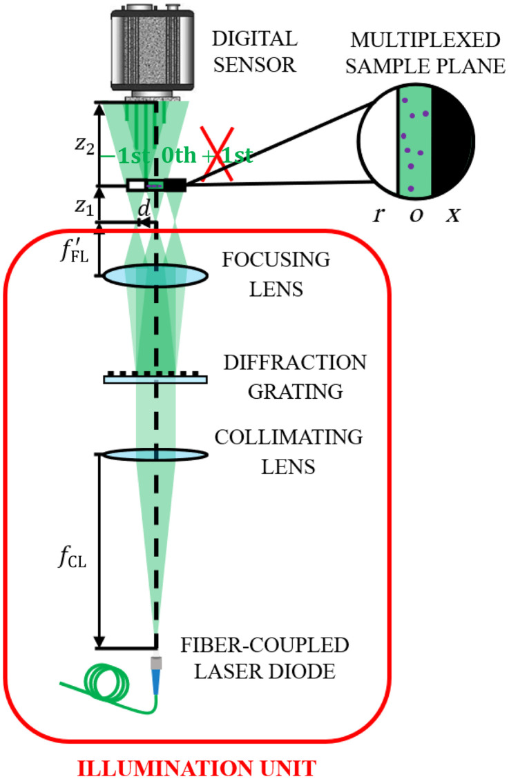
Aim: In addition to the abovementioned advantages, in-line arrangements for DLHM also include the limitation of the twin-image presence, which can restrict accurate QPI. We therefore propose a compact lensless common-path interferometric off-axis approach that is capable of quantitative imaging of fast-moving biological specimens, such as living cells in flow.

Approach: We suggest lensless spatially multiplexed interferometric microscopy (LESSMIM) as a lens-free variant of the previously reported spatially multiplexed interferometric microscopy (SMIM) concept. LESSMIM comprises a common-path interferometric architecture that is based on a single diffraction grating to achieve digital off-axis holography. From a series of single-shot off-axis holograms, twin-image free and time-resolved QPI is achieved by commonly used methods for Fourier filtering-based reconstruction, aberration compensation, and numerical propagation.

Results: Initially, the LESSMIM concept is experimentally demonstrated by results from a resolution test chart and investigations on temporal stability. Then, the accuracy of QPI and capabilities for imaging of living adherent cell cultures is characterized. Finally, utilizing a microfluidic channel, the cytometry of suspended cells in flow is evaluated.

Conclusions: LESSMIM overcomes several limitations of in-line DLHM and provides fast time-resolved QPI in a compact optical arrangement. In summary, LESSMIM represents a promising technique with potential biomedical applications for fast imaging such as in imaging flow cytometry or sperm cell analysis.

Keywords: digital holographic microscopy; digital lensless holographic microscopy; label-free imaging; off-axis lensless holography; phase retrieval; quantitative phase imaging; spatially multiplexed interferometric microscopy.


Figures

Fig. 1
Fig. 1
Scheme of the LESSMIM layout consisting of an illumination unit and a digital recording sensor. fCL is the focal length of the collimating lens for object illumination with laser light, fFL is the focal length of the focusing lens for the creation of three-point sources, z1 is the distance between the point light sources and sample, z2 is the distance between the sample and digital sensor, o is the object region, r is the reference region, and x is the blocking region.
Fig. 2
Fig. 2
Illustration of the LESSMIM working principle for the example of a USAF resolution test target: (a) recorded off-axis hologram; (b) Fourier transformation of panel (a) with the filtered spectral region (marked with a dotted red circle); (c) amplitude and (d) phase reconstructions after Fourier filtering application; (e) amplitude and (f) phase reconstructions of the reference hologram; (g) amplitude and (h) phase images after subtraction of the amplitude and phase distributions retrieved from the reference hologram; (i) amplitude and (j) phase images after numerical propagation to the focus plane; and (k) amplitude and (l) phase images recovered by conventional in-line DLHM without a separate reference wave for direct comparison; (m) color-coded magnified images of regions in panels (i)–(l) containing the smallest elements of the resolution target; and (n) intensity profiles along blue and red lines in panels (i) and (k), respectively. The scale bar in panel (a) corresponds to a length of 20  μm.
Fig. 3
Fig. 3
Temporal phase stability of LESSMIM during a period of 300 s within an ROI of 300×300  pixels utilizing a blank microscope slide as the sample location (hologram acquisition rate of 1 Hz). (a) temporal (t) development of the averaged phase Δφ(t)¯; (b) spatial distribution of the standard deviation σt(m,n) of the phase differences per pixel determined from the entire stack of 300 QPI images; and (c) histogram of the σt from the data in panel (b). The mean value σt¯=0.07  rad quantifies a high temporal stability of the setup. The scale bar in panel (b) corresponds to 20  μm.
Fig. 4
Fig. 4
Validation of QPI image retrieval with LESSMIM by analysis of PMMA microspheres in a glycerol/water mixture (90%/10%). (a) Recorded off-axis hologram, (b) reconstructed numerically focused QPI image, (c) gray level coded pseudo-3D representation of thickness distribution computed from phase data in panel (b), and (d) thickness profile along the dotted blue line in panel (b). Yellow and white scale bars in panels (a) and (b) correspond to 100 and 20  μm, respectively.
Fig. 5
Fig. 5
Evaluation of LESSMIM for QPI of living adherent pancreatic tumor cells (PaTu 8988T). Rows (1)–(4): ROIs containing cells with various morphologies and thicknesses. First column (a1–a4): recorded off-axis holograms; second column (b1–b4): reconstructed focused QPI images; third column (c1–c4): gray level coded pseudo-3D plots of the cell thickness distributions calculated from panels (b1–b4); and fourth column (d1–d4): thickness profile along blue dotted lines marked in panels (b1–b4). Scale bars in panels (a1–a4) correspond to 20  μm.
Fig. 6
Fig. 6
Evaluation of LESSMIM for IFC by investigations on living Patu 8988T cells in a microfluidic chip with hydrodynamic focusing capabilities. (a) Scheme of the utilized microfluidic chip with the enlarged region (green-framed rectangular) considered for lensless holographic imaging by spatial multiplexing (x is the blocking region, o is the object region, and r is the reference region). (b)–(d) Representative images of a recorded movie of 5 s that was recorded at a hologram acquisition rate of 40 fps with an exposure time of 2 ms (Video 1). (b) Representative off-axis hologram (the yellow arrow indicates the direction of the sample fluid stream); (c) focused QPI image reconstructed from panel (b); and (d) pseudo-3D thickness distribution computed from panel (c), with green outlines being the area retrieved by segmentation and indicating the cell boundaries. (e)–(f) Simultaneous visualization of all detected cells in the recorded QPI image stack achieved by maximum intensity projection (MIP); (e) MIP QPI image; and (f) corresponding pseudo-3D thickness representation with outlined cell boundaries. (Video 1, MOV, 7.67 MB [URL: https://doi.org/10.1117/1.JBO.29.S2.S22715.s1]).
Fig. 7
Fig. 7
Determination of biophysical parameters from 300 individually analyzed suspended pancreatic tumor cells (Patu 8988T) in flow as illustrated in Fig. 6. (a) Scatter plot of the integral cellular RI n versus the projected cell radius R and (b) the relative frequency histogram of the dry mass DM.

Similar articles

Cited by

References

    1. Kim M. K., Digital holographic microscopy. Principles, techniques, and applications, Springer US; (2011).
    1. Park Y. K., Depeursinge C., Popescu G., “Quantitative phase imaging in biomedicine,” Nat. Photonics 12(10), 578–589 (2018).NPAHBY10.1038/s41566-018-0253-x - DOI
    1. Kemper B., et al. , “Label-free quantitative in vitro live cell imaging with digital holographic microscopy,” in Bioanalytical Reviews, Wegener J., Ed., Vol. 2, Springer, Cham: (2019).
    1. Zernike F., “Phase contrast, a new method for the microscopic observation of transparent objects,” Physica 9(7), 686–698 (1942).10.1016/S0031-8914(42)80035-X - DOI
    1. Allen R., David G., Nomarski G., “The Zeiss-Nomarski differential interference equipment for transmitted-light microscopy,” Zeitschrift fur wissenschaftliche Mikroskopie 69(4), 193–221 (1969). - PubMed

Publication types

MeSH terms