. 2024 Dec 3;23(12):1854-1865.
doi: 10.1158/1535-7163.MCT-23-0833.

Trametinib Potentiates Anti-PD-1 Efficacy in Tumors Established from Chemotherapy-Primed Pancreatic Cancer Cells

Affiliations

Trametinib Potentiates Anti-PD-1 Efficacy in Tumors Established from Chemotherapy-Primed Pancreatic Cancer Cells

Thao D Pham et al. Mol Cancer Ther. .

Abstract

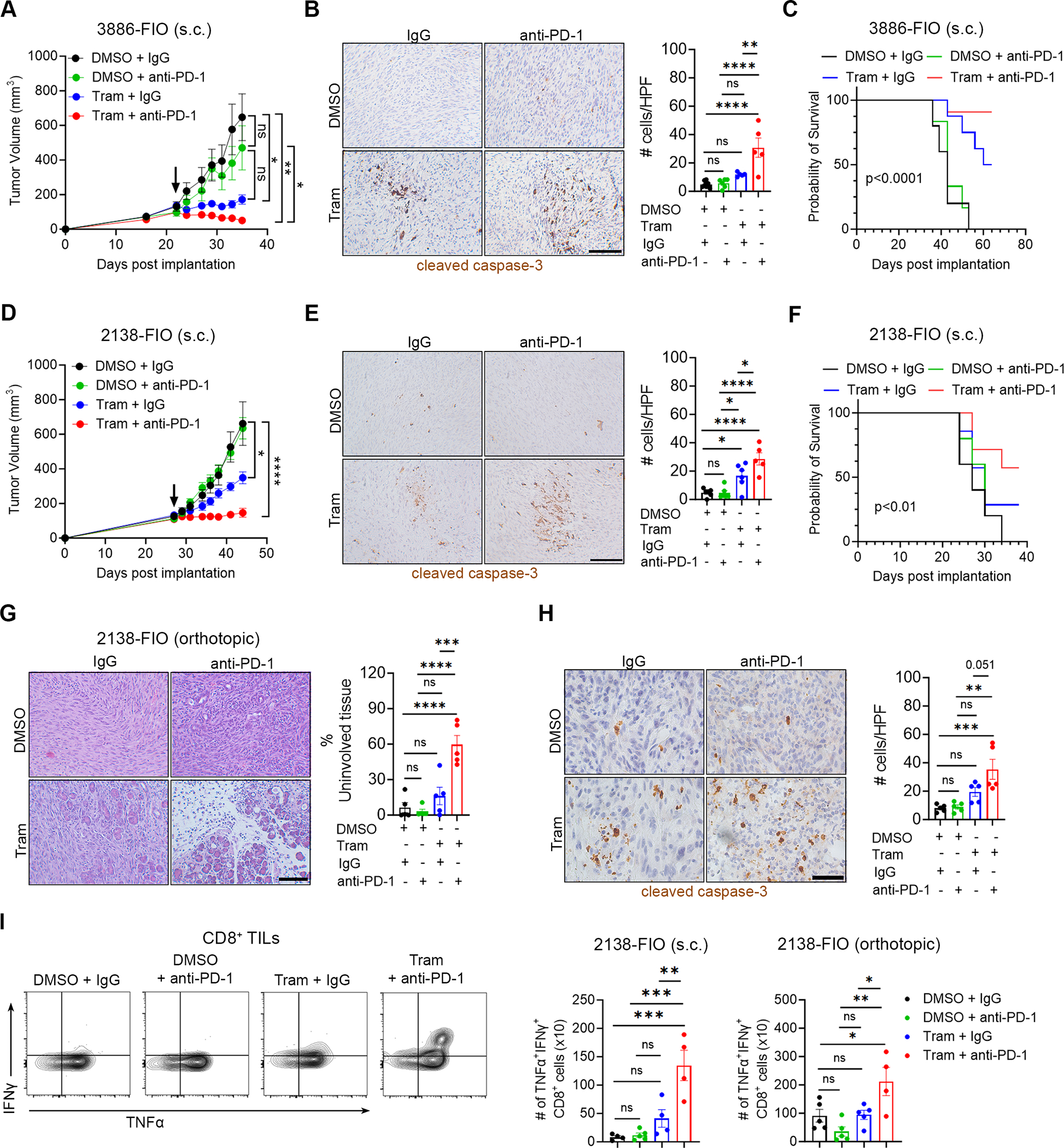
Despite advances in immune checkpoint inhibitors, chemotherapy remains the standard therapy for patients with pancreatic ductal adenocarcinoma (PDAC). As the combinations of chemotherapy, including the FOLFIRINOX [5-fluorouracil, F; irinotecan, I; and oxaliplatin, O (FIO)] regimen, and immune checkpoint inhibitors have failed to demonstrate clinical benefit in patients with metastatic PDAC tumors, there is increasing interest in identifying therapeutic approaches to potentiate ICI efficacy in patients with PDAC. In this study, we report that neoadjuvant FOLFIRINOX-treated human PDAC tumors exhibit increased MEK/ERK activation. We also show elevated MEK/ERK signaling in ex vivo PDAC slice cultures and cell lines treated with a combination of FIO. In addition, we find that the KPC-FIO cells, established from repeated treatment of mouse PDAC cell lines with six to eight cycles of FIO, display enhanced ERK phosphorylation and demonstrate increased sensitivity to MEK inhibition in vitro and in vivo. Significantly, the KPC-FIO cells develop tumors with a proinflammatory immune profile similar to human PDAC tumors after neoadjuvant FOLFIRINOX treatment. Furthermore, we found that the MEK inhibitor trametinib enables additional infiltration of highly functional CD8+ T cells into the KPC-FIO tumors and potentiates the efficacy of anti-PD-1 antibody in syngeneic mouse models. Our findings provide a rationale for combining trametinib and anti-PD-1 antibodies in patients with PDAC after neoadjuvant or short-term FOLFIRINOX treatment to achieve effective antitumor responses.


Conflict of interest statement

The authors declare no potential conflicts of interest

Figures

Fig. 1.
Fig. 1.. Combination of 5FU, irinotecan, and oxaliplatin increases ERK phosphorylation in human and mouse pancreatic cancer models.
A. Schematic diagram illustrating neoadjuvant treatment with FOLFIRINOX in patients with localized pancreatic ductal adenocarcinoma (PDAC). B. Tumors from PDAC patients receiving neoadjuvant FOLFIRINOX and patients undergoing up-front surgery were co-stained for p-ERKThr202/Tyr204 and cytokeratin 19 (CK19) and counterstained with DAPI. Scale bar 150 μm. The number of p-ERK+ cells as a percentage of DAPI+ cells was quantified by ImageJ and analyzed by GraphPad. Error bar ± SEM. Unpaired t-test. **, p<0.01. C. Schematic diagram depicting how slice cultures are generated. 6mm punch biopsies from mouse and human PDAC tumors are sectioned by a vibratome to 250 μm thick slices and treated with chemotherapy. After 72–96 hours, slices are collected and analyzed by H&E and immunofluorescent (IF) staining. D, E. Slice cultures from human PDAC tumors (D) and a mouse KPC tumor (E) were treated with either vehicle control (Ctrl) or a combination of 5FU (F, 4 μM), SN-38 (I, 12.5 nM), and oxaliplatin (O, 0.5 μM) (FIO). After 72–96 hours, slices were collected and analyzed for histology by H&E staining and p-ERKThr202/Tyr204/CK19/DAPI by immunofluorescent staining. Scale bar 75 μm. The number of p-ERK+ cells as a percentage of DAPI+ cells was quantified by ImageJ, normalized to the vehicle-treated samples, and analyzed by GraphPad. Paired t-test. *, p<0.05. F. Mouse KPC cell lines (2138, 3886, 1245, 3213) were treated with a combination of 5FU (F, 4 μM), SN-38 (I, 12.5 nM), and oxaliplatin (O, 0.5 μM) (FIO) for 24 hours and analyzed for ERKThr202/Tyr204 phosphorylation and total ERK by western blotting with HSP90 as loading control. Blots are representative of at least three biological replicates.
Fig. 2.
Fig. 2.. KPC-FIO cells demonstrate increased ERK phosphorylation in vitro and in vivo.
A. Schematic diagram depicting how KPC-FIO cells are generated. Parental KPC cells are cultured in the presence of 4 μM 5FU, 12.5 nM SN-38 (the active metabolite of irinotecan), and 0.5 μM oxaliplatin for 3 days, then allowed to recover in drug-free media for 4 days. KPC-FIO cells are obtained after 6–8 cycles of treatment. B. Phase (top) and actin filament (bottom) pictures of the parental KPC (2138, 3886, 3213, 1245) and the corresponding KPC-FIO cells. Cells were stained for actin filaments using phalloidin and counterstained with DAPI following protocol described in Materials & Methods. Scale bar, 50 μm. C. Gene set enrichment analysis (GSEA) comparing 3213 (n = 3) and 3213-FIO (n = 3) cells for negative regulators of the MAPK kinase pathway. NES, normalized enrichment score. D. The KPC and KPC-FIO cells were analyzed for p-ERK1/2Thr202/Tyr204 and total ERK1/2 expression by western blotting with HSP90 as loading control. Blots are representative of at least three biological replicates. E. 3213, 3213-FIO, 2138, 2138-FIO, 3886, and 3886-FIO cells were subcutaneously implanted in C57BL/6 mice and allowed to form established tumors. The resulting tumors were immunofluorescence co-stained for p-ERKThr202/Tyr204 and CK19 and counterstained with DAPI. Scale bar 75 μm. The number of p-ERK+ cells as a percentage of DAPI+ cells were quantified by ImageJ and analyzed by GraphPad. Error bar ± SEM. Unpaired t-test. ***, p<0.001 ****, p<0.0001.
Fig. 3.
Fig. 3.. Trametinib induces apoptosis in the KPC-FIO cells and tumors.
A. The KPC and KPC-FIO cells were treated with DMSO, U0126 (2 μM), Binimetinib (2 μM, Bini), Cobimetinib (2 μM, Cobi), Selumetinib (2 μM, Selu), or Trametinib (2 μM, Tram) for 72 hours. The effects of different MEK inhibitors on cell proliferation were analyzed by WST-1 assay. Error bars ± SEM. *, p<0.05 **, p<0.01 ***, p<0.001 ****, p<0.0001. B. The KPC and KPC-FIO cells were treated with either vehicle control or Trametinib at the indicated doses for 48 hours and evaluated by Annexin V/PI staining for cell death. Representative flow blots were shown for 0.2 μM Trametinib. Error bars ± SEM. *, p<0.05 **, p<0.01 ***, p<0.001 ****, p<0.0001 ns, non-significant. C-F. Mice with established 2138 (C) and 2138-FIO (E) tumors were treated with DMSO as vehicle control or Trametinib (1 mg/kg, daily). Arrow indicates start of treatment. Tumor size was measured 2–3 times/week by caliper, and tumor volume was calculated using the formula V = (W2 × L)/2. Error bar ± SEM from 8–10 tumors. Unpaired t-test. *, p<0.05 **, p<0.01 ***, p<0.001 ****, p<0.0001 ns, non-significant. At the end, tumors were collected and co-stained for p-ERKThr202/Tyr204 and CK19 and counterstained with DAPI. Scale bar 75 μm. The DMSO- or Trametinib-treated 2138 (D) and 2138-FIO (F) tumors at endpoint were evaluated for proliferation and cell death by immunohistochemistry staining of Ki67 and cleaved caspase-3 (CC3), respectively. Scale bar, 100 μm. The number of positive cells was analyzed by ImageJ. Unpaired t-test. *, p<0.05 **, p<0.01 ***, p<0.001 ns, non-significant.
Fig. 4.
Fig. 4.. Tumors established from KPC-FIO cells share similar clinical features with neoadjuvant FOLFIRINOX-treated human tumors.
A. Tumors from neoadjuvant FOLFIRINOX-treated PDAC patients were stained by immunohistochemistry for CD8, CD68, CD206, and Arginase-1, and compared to tumors from patient undergoing up-front surgery. Scale bar 100 μm. The number of positive cells was analyzed by ImageJ. Error bars ± SEM. Unpaired t-test. *, p<0.05. B-D. 2138, 2138-FIO (B), 3886, 3886-FIO (C), and 3213, 3213-FIO (D) cells were subcutaneously implanted in C57BL/6 mice and allowed to form established tumors. At endpoint, tumors were collected and stained by immunohistochemistry for CD8, F4/80, CD206, and Arginase-1. Scale bar 100 μm. The number of positive cells was analyzed by ImageJ. Error bars ± SEM. Unpaired t-test. *, p<0.05 **, p<0.01 ****, p<0.0001 ns, non-significant. E-G. Established 2138, 2138-FIO (E), 3886, 3886-FIO (F), and 3213, 3213-FIO (G) tumors were treated with an anti-PD-1 antibody (250 μg, 3 times/week) or a control isotype-matched IgG (250 μg, 3 times/week). Arrow indicates start of treatment. Tumor size was measured 2–3 times/week by caliper, and tumor volume was calculated using the formula V = (W2 × L)/2. Error bars ± SEM from 4–8 tumors.
Fig. 5.
Fig. 5.. Trametinib enhances infiltration by functional CD8+ T cells into the KPC-FIO tumors.
A-D. 2138 (A), 2138-FIO (B), 3886 (C), and 3886-FIO (D) cells were subcutaneously implanted in C57BL/6 mice and allowed to form tumors. Mice with established tumors were treated with DMSO as vehicle control or Trametinib (1 mg/kg, daily) for 2 weeks. DMSO or Trametinib-treated tumors at endpoint were processed and analyzed by flow cytometry for the frequency of CD4+ T cells (CD45+CD4+), CD8+ T cells (CD45+CD8+), and CD8+ T cell functional markers (IFNγ, Granzyme B (GzmB), TNFα, and Perforin-1 (Prf1)). Error bars ± SEM. Unpaired t-test. *, p<0.05 **, p<0.01 ***, p<0.001 ns, non-significant. All cells were gated from single live cells.
Fig. 6.
Fig. 6.. Trametinib sensitizes KPC-FIO tumors to anti-PD-1 treatment.
A-F. Mice with established subcutaneous (s.c.) 3886-FIO (A) and 2138-FIO (D) tumors were randomized and treated with Trametinib (Tram, 1 mg/kg, daily) or vehicle control (DMSO) in combination with an anti-PD-1 antibody (250 μg, 3 times/week) or a control isotype-matched IgG antibody (250 μg, 3 times/week). Arrow indicates the start of treatment. Tumor size was measured 2–3 times/week by caliper, and tumor volume was calculated using the formula V = (W2 × L)/2. Treated 3886-FIO (B) and 2138-FIO (E) tumors at endpoint were evaluated for cell death by immunohistochemistry staining of cleaved caspase-3. Scale bar, 100 μm. The number of positive cells was analyzed by ImageJ. The survival of tumor-bearing mice was evaluated and analyzed by Log-rank (Mantel-Cox) test (C, F). G, H. Mice with established orthotopic 2138-FIO tumors were randomized and treated with Trametinib (Tram, 1 mg/kg, daily) or DMSO as vehicle control in combination with an anti-PD-1 antibody (250 μg, 3 times/week) or a control isotype-matched IgG antibody (250 μg, 3 times/week). After 2 weeks, tumors were collected and evaluated for the extent of uninvolved pancreatic tissues by H&E staining (G) and cell death by cleaved caspase-3 staining (H). Scale bar, 100 μm. The number of positive cells was analyzed by ImageJ. I. Subcutaneous (s.c.) and orthotopic 2138-FIO tumors at study endpoint were evaluated for the absolute number of CD8+ cells (per 100,000 live cells) simultaneously expressing the activation markers IFNγ and TNFα. Error bars ± SEM. All analyses were performed using one-way ANOVA, followed by Tukey’s multiple comparison test. *, p<0.05 **, p<0.01 ***, p<0.001 ****, p<0.0001 ns, non-significant.

References

    1. Rahib L, Smith BD, Aizenberg R, Rosenzweig AB, Fleshman JM, and Matrisian LM (2014). Projecting cancer incidence and deaths to 2030: the unexpected burden of thyroid, liver, and pancreas cancers in the United States. Cancer Res 74, 2913–2921. 10.1158/0008-5472.CAN-14-0155. - DOI - PubMed
    1. Siegel RL, Giaquinto AN, and Jemal A (2024). Cancer statistics, 2024. CA Cancer J Clin 74, 12–49. 10.3322/caac.21820. - DOI - PubMed
    1. Conroy T, Desseigne F, Ychou M, Bouche O, Guimbaud R, Becouarn Y, Adenis A, Raoul JL, Gourgou-Bourgade S, de la Fouchardiere C, et al. (2011). FOLFIRINOX versus gemcitabine for metastatic pancreatic cancer. N Engl J Med 364, 1817–1825. 10.1056/NEJMoa1011923. - DOI - PubMed
    1. Ghaneh P, Palmer D, Cicconi S, Jackson R, Halloran CM, Rawcliffe C, Sripadam R, Mukherjee S, Soonawalla Z, Wadsley J, et al. (2023). Immediate surgery compared with short-course neoadjuvant gemcitabine plus capecitabine, FOLFIRINOX, or chemoradiotherapy in patients with borderline resectable pancreatic cancer (ESPAC5): a four-arm, multicentre, randomised, phase 2 trial. Lancet Gastroenterol 8, 157–168. 10.1016/S2468-1253(22)00348-X. - DOI - PubMed
    1. Katz MH, Shi Q, Ahmad SA, Herman JM, Marsh Rde W, Collisson E, Schwartz L, Frankel W, Martin R, Conway W, et al. (2016). Preoperative Modified FOLFIRINOX Treatment Followed by Capecitabine-Based Chemoradiation for Borderline Resectable Pancreatic Cancer: Alliance for Clinical Trials in Oncology Trial A021101. JAMA Surg 151, e161137. 10.1001/jamasurg.2016.1137. - DOI - PMC - PubMed

MeSH terms