. 2024 Oct 14;52(18):10986-10998.
doi: 10.1093/nar/gkae708.

Dispensability of HPF1 for cellular removal of DNA single-strand breaks

Affiliations

Dispensability of HPF1 for cellular removal of DNA single-strand breaks

Kristyna Hrychova et al. Nucleic Acids Res. .

Abstract

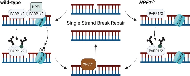
In response to DNA damage, the histone PARylation factor 1 (HPF1) regulates PARP1/2 activity, facilitating serine ADP-ribosylation of chromatin-associated factors. While PARP1/2 are known for their role in DNA single-strand break repair (SSBR), the significance of HPF1 in this process remains unclear. Here, we investigated the impact of HPF1 deficiency on cellular survival and SSBR following exposure to various genotoxins. We found that HPF1 loss did not generally increase cellular sensitivity to agents that typically induce DNA single-strand breaks (SSBs) repaired by PARP1. SSBR kinetics in HPF1-deficient cells were largely unaffected, though its absence partially influenced the accumulation of SSB intermediates after exposure to specific genotoxins in certain cell lines, likely due to altered ADP-ribosylation of chromatin. Despite reduced serine mono-ADP-ribosylation, HPF1-deficient cells maintained robust poly-ADP-ribosylation at SSB sites, possibly reflecting PARP1 auto-poly-ADP-ribosylation at non-serine residues. Notably, poly-ADP-ribose chains were sufficient to recruit the DNA repair factor XRCC1, which may explain the relatively normal SSBR capacity in HPF1-deficient cells. These findings suggest that HPF1 and histone serine ADP-ribosylation are largely dispensable for PARP1-dependent SSBR in response to genotoxic stress, highlighting the complexity of mechanisms that maintain genomic stability and chromatin remodeling.


Figures

Graphical Abstract
Graphical Abstract
Figure 1.
Figure 1.
HPF1 is largely dispensable for cell survival in response to DNA single-strand breaks. (A) Immunofluorescence analysis of endogenous mono-ADP-ribosylation levels detected by a specific mono-ADP-ribose binding reagent (MAR 205) in U2OS (II) wild-type, HPF1−/− (II-cl.1), ARH3−/−/HPF1−/− (clone #D) and ARH3−/− (clone #48) cells. Representative ScanR images and quantifications are shown, RFU - relative fluorescence units. Data are the mean (±SD) of three independent experiments. Statistical analysis (one-way analysis of variance) is shown (ns – not significant, ****P < 0.0001). (BC) Clonogenic survival assay in U2OS (I) wild-type, HPF1−/− (clone I-#5 and I-#8), PARP1−/−/PARP2−/− (clone #5) and U2OS (II) wild-type and HPF1−/− (II-cl.1) cells in response to treatment with indicated doses of CPT and MMS for 15 min at RT. Data represent the mean (±SD) of three independent experiments. Statistical analysis (two-way analysis of variance) is shown (ns – not significant, **P < 0.01, ***P < 0.001, ****P < 0.0001). (D, E) Clonogenic survival assay in RPE-1 wild-type, HPF1−/− (clone #5 and #10), PARP1−/−/PARP2−/− (clone #E6), and PARP1−/− (clone #G7), PARP2−/− (clone #A1), HPF1−/−/PARP1−/− (clone #14 and #21) and HPF1−/−/PARP2−/− (clone #3 and #7) cells in response to treatment with indicated doses of CPT and MMS for 15 minutes at RT. Data represent the mean (±SD) of three independent experiments. Statistical analysis (two-way analysis of variance) is shown (ns – not significant, *P < 0.05, **P < 0.01, ***P < 0.001, ****P < 0.0001).
Figure 2.
Figure 2.
Impact of HPF1 deficiency on DNA single-strand break repair. (A) DNA strand breaks quantified by alkaline comet assays (Methods II) in U2OS (I) wild-type, HPF1−/− (clone I-#5 and I-#8), and U2OS (II) wild-type and HPF1−/− (II-cl.1), cells before and after 100 μM H2O2 treatment in serum-free media for 10 min on ice, followed by incubation at 37°C in full media (release time), in the presence or absence of 10 μM PARPi. The individual comet tail moments of cells combined from three to four independent experiments are plotted (the upper chart). A minimum of 50 cells were analysed per sample in each of the experiments. The normalized data are shown (the lower chart) and represent the relative mean (±SD) of three to four independent experiments. Statistical analysis (two-way analysis of variance) is shown (ns, not significant). (B) Alkaline comet assay analysis (Methods I) in RPE-1 wild-type, HPF1−/− (clone #5 and #10), and PARP1−/−/PARP2−/− (clone #E6) cells before and after 50 μM H2O2 treatment in serum-free media for 10 min on ice, followed by incubation at 37°C in full media (release time). The individual comet tail moments of cells combined from three independent experiments are plotted (the upper chart). A minimum of 50 cells were analysed per sample in each of the experiments. The normalized data are shown (the lower chart) and represent the relative mean (±SD) of three independent experiments. Statistical analysis (two-way analysis of variance) is shown (**P < 0.01, ****P < 0.0001). (C, D) Alkaline comet assay analysis (Methods II) in U2OS (C) or RPE-1 (D) clones, as indicated, before or after the treatment with 0.9 mM MMS for 15 min at 37°C, followed by incubation at 37°C after MMS wash (release time). The individual comet tail moments of cells combined from three independent experiments are plotted (the upper charts). A minimum of 50 cells were analysed per sample in each of the experiments. The normalized data are shown (the lower charts) and represent the relative mean (±SD) of three independent experiments. Statistical analysis (two-way analysis of variance) is shown (ns – not significant, *P < 0.05, **P < 0.01, ****P < 0.0001).
Figure 3.
Figure 3.
PARP1-dependent ADP-ribosylation following DNA damage in both wild-type and HPF1-deficient cells. (A) ADP-ribosylation levels in RPE-1 wild-type, PARP1−/− (clone #G7), PARP2−/− (clone #A1), and PARP1−/−/PARP2−/− (clone #E6) cells after treatment with 2 mM H2O2 for 20 min or 0.9 mM MMS for 1 hour in full media at 37°C detected by western blotting using the iAf1521 reagent (MAR/PAR). (B) ADP-ribosylation levels in RPE-1 wild-type, PARP1−/− (clone #G7), PARP2−/− (clone #A1), PARP1−/−/PARP2−/− (clone #E6) and HPF1−/− (clone #5) cells before and after treatment with 2 mM H2O2 for 20 min in full media at 37°C detected by western blotting using a specific anti-mono-ADP-ribose binding reagent (MAR 647) or anti-poly-ADP-ribose antibody (PAR). The asterisk denotes a nonspecific band, resulting from a cross-reaction with a component from the serum. (C) ADP-ribosylation levels in HPF1−/− (clone #5), HPF1−/−/PARP1−/− (clone #14 and #21), and HPF1−/−/PARP2−/− (clone #3 and #7) cells after treatment with 2 mM H2O2 for 20 min or 0.9 mM MMS for 1 hour in full media at 37°C detected by western blotting using a specific anti-poly-ADP-ribose antibody (PAR).
Figure 4.
Figure 4.
The elongated poly-ADP-ribose chains in HPF1-deficient cells after DNA damage. (A) Poly-ADP-ribosylation in RPE-1 wild-type and HPF1−/− (clone #5 and #10) cells after 100 μM H2O2 treatment in serum-free media for 10 min on ice, followed by incubation at 37°C in full media (release time) detected by western blotting using a specific anti-poly-ADP-ribose antibody (PAR). The asterisk denotes a nonspecific band, resulting from a cross-reaction with a component from the serum. (B) Poly-ADP-ribosylation in RPE-1 wild-type and HPF1−/− (clone #5 and #10) cells after treatment with 0.9 mM MMS for 30 and 60 min at 37°C detected by western blotting using a specific anti-poly-ADP-ribose antibody (PAR). The asterisk denotes a nonspecific band, resulting from a cross-reaction with a component from the serum. (CD) Immunofluorescence analysis of ADP-ribosylation levels detected by the specific mono-ADP-ribosylation (MAR 205) binding reagent or the specific poly-ADP-ribosylation (PAR) antibody after detergent-extraction in untreated RPE-1 wild-type, HPF1−/− (clone #5 and #10), and PARP1−/−/PARP2−/− (clone #E6) cells and after treatment with 2 mM H2O2 in full media for 20 min at 37°C. Data represents the mean (±SD) of four independent experiments, RFU—relative fluorescence units. Statistical analysis (one-way analysis of variance) is shown (**P < 0.01, ****P < 0.0001). The corresponding representative ScanR images for immunofluorescence data are shown. (E) Immunofluorescence analysis of ADP-ribosylation levels detected by the specific poly-ADP-ribosylation (PAR) antibody after detergent-extraction in untreated RPE-1 wild-type and HPF1−/− (clone #5 and #10) cells and after treatment with 0.9 mM MMS for 1 h at 37°C. Data represents the mean (±SD) of three independent experiments, RFU – relative fluorescence units. Statistical analysis (one-way analysis of variance) is shown (ns, not significant). The corresponding representative ScanR images for immunofluorescence data are shown.
Figure 5.
Figure 5.
XRCC1 recruitment into chromatin following DNA damage remains unaffected in HPF1-deficient cells. (A) U2OS (I) wild-type, HPF1−/− (clone I-#5 and I-#8), PARP1−/−/PARP2−/− (clone #5), U2OS (II) wild-type, and HPF1−/− (II-cl.1) cells were transiently transfected with mRFP-XRCC1 and microirradiated with a 405 nm UV-laser. Representative images captured pre-irradiation, and at one and five minutes post-irradiation, and quantifications are shown. Data represents the mean percentage (±SD) of transfected cells with XRCC1 recruitment to DNA damage sites, averaged over three independent experiments. Statistical analysis (one-way analysis of variance) is shown (ns, not significant, *P < 0.05, ****P < 0.0001). A minimum of 17 transfected cells were analysed per sample in each experiment. (B) Immunofluorescence analysis of chromatin-bound nuclear XRCC1 after detergent-extraction in RPE-1 wild-type, HPF1−/− (clone #5 and #10), and XRCC1−/− (clone #3) cells untreated or treated with indicated doses of H2O2 in serum-free media for 10 min on ice. Representative ScanR images and quantifications are shown, RFU—relative fluorescence units. Data are the mean (±SD) of three independent experiments. Statistical analysis (one-way analysis of variance) is shown (ns, not significant, ****P < 0.0001). (C) Levels of HPF1 and XRCC1 in whole cell (WC) extracts and chromatin-containing fractionations from RPE-1 wild-type, HPF1−/− (clone #5 and #10) cells before and following treatment with 2 mM H2O2 in full media for 20 min at 37°C. Normalized chromatin bound XRCC1 protein levels, quantified from the western blots, represent the mean (±SD) from four independent experiments. Statistical analysis (one-way analysis of variance) is shown (ns, not significant).

Similar articles

Cited by

References

    1. Lindahl T. Instability and decay of the primary structure of DNA. Nature. 1993; 362:709–715. - PubMed
    1. Caldecott K.W. DNA single-strand break repair and human genetic disease. Trends Cell Biol. 2022; 32:733–745. - PubMed
    1. Pommier Y., Sun Y., Huang S.N., Nitiss J.L.. Roles of eukaryotic topoisomerases in transcription, replication and genomic stability. Nat. Rev. Mol. Cell Biol. 2016; 17:703–721. - PMC - PubMed
    1. Demple B., Harrison L.. Repair of oxidative damage to DNA: enzymology and biology. Annu. Rev. Biochem. 1994; 63:915–948. - PubMed
    1. Caldecott K.W. Mammalian DNA base excision repair: dancing in the moonlight. DNA Repair (Amst.). 2020; 93:102921. - PubMed

MeSH terms